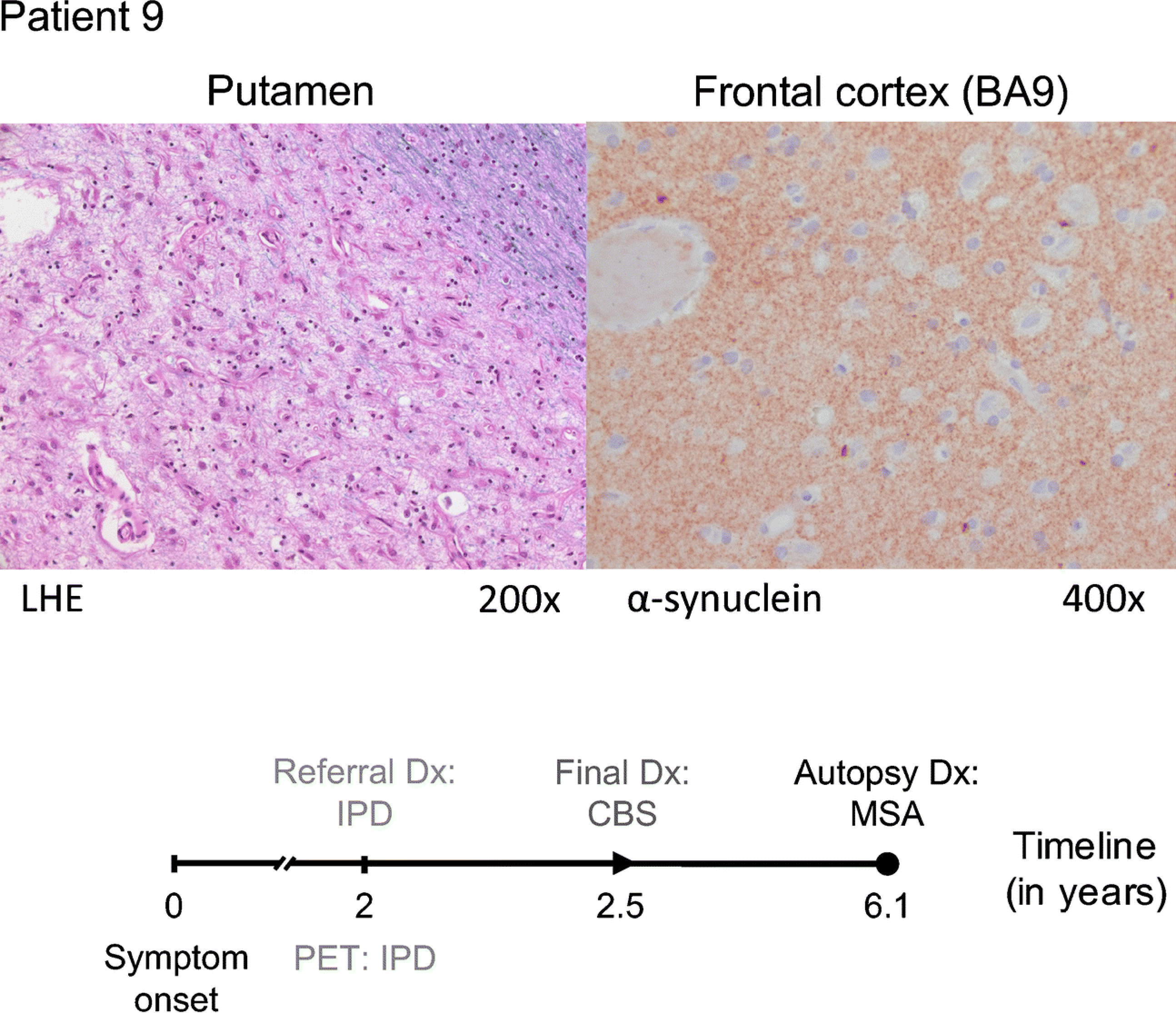
Fig. 3. A challenging case of pathology-proven multiple system atrophy (MSA).

Patient 9 (64-year old female, symptom duration 2 years) was referred for FDG PET because of an uncertain clinical diagnosis with a suspicion of IPD or CBS. The automated imaging algorithm classified the patient as IPD (83.9% likelihood). After 6 months of additional clinical follow-up, the clinical diagnosis was revised to CBS. Autopsy performed four years later revealed changes consistent with MSA: severe neuronal loss and reactive gliosis in the putamen (LHE, 200×; left), pons, substantia nigra, and cerebellum. α-synuclein-labeled glial cytoplasmic inclusions were found in the frontal cortex (α-synuclein antibody, 400×; right), the striatum, the pons, amygdala, and medulla oblongata.
