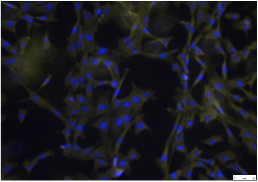
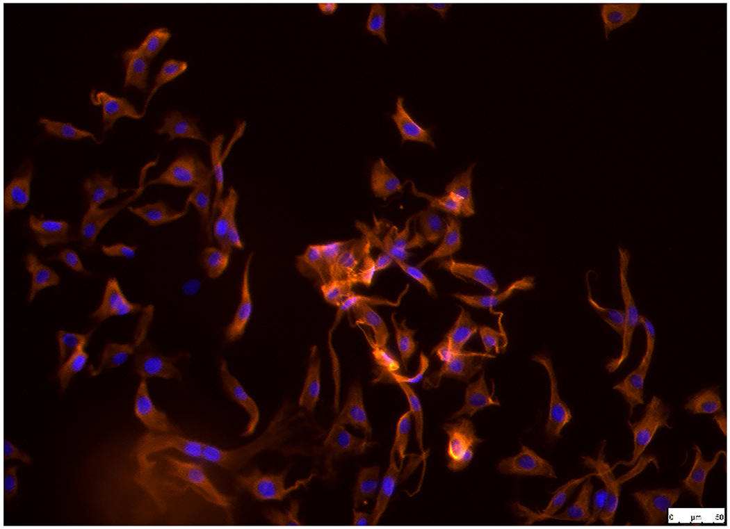
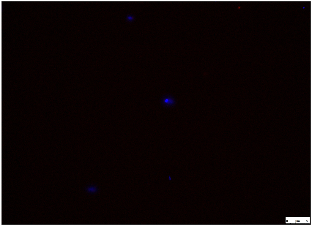
Figure 1.


Establishment and characterization of pAVICs. Cells were positive for (A) α-SMA and (B) vimentin in pAVICs. (C) BSA (1%) was used as a negative control in order to exclude non-specific fluorescence staining. Scale bar, 50 μm. α-SMA, a-smooth muscle actin; pAVICs, porcine aortic valve interstitial cells.
