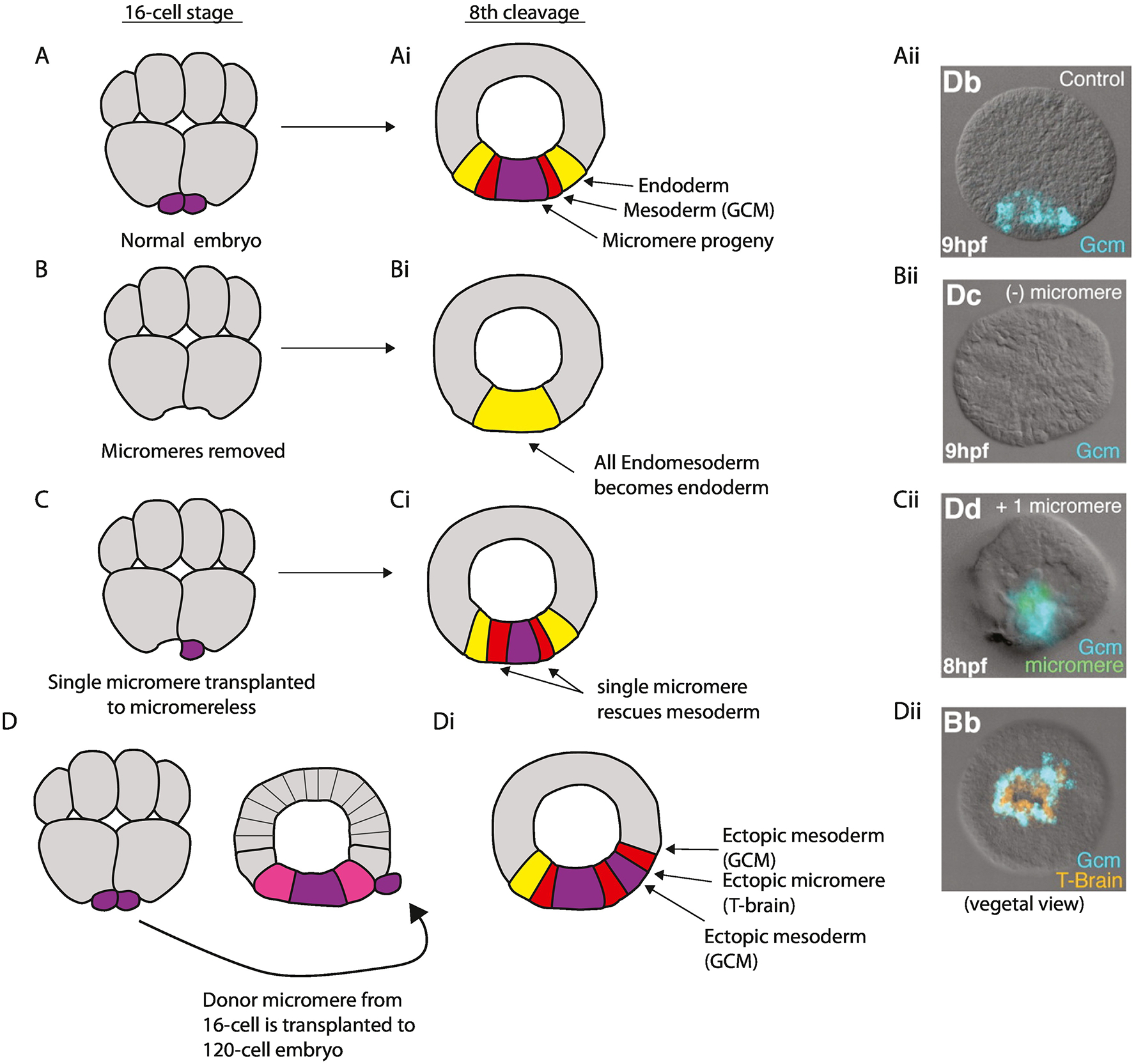
Figure 3:

Delta-Notch signaling from the micromeres establishes mesoderm. Micromeres first appear at the 16-cell stage (A), activate delta expression at the 32-cell stage and begin expressing Delta at the 60-cell stage (Croce and McClay, 2010). (Ai) This signaling induces mesoderm gene expression in the adjacent endomesoderm (Veg2) cell layer (Ransick et al., 2002; Croce and McClay, 2010; Peter and Davidson, 2010). (Aii) Gcm, a mesoderm marker in the endomesoderm, is expressed at hatched blastula (HB) (reproduced from Croce and McClay, 2010). (B) If the micromeres are surgically removed at the 16-cell stage all endomesoderm is specified as endoderm only (Bi) because Delta is absent. (Bii) Consequently gcm is not expressed. (C) A single labeled micromere reintroduced at 16-cell stage in an otherwise micromere deficient embryo is sufficient to rescue mesoderm development and gcm expression (Ci, Cii). (D) If a micromere from a 16-cell stage embryo is transplanted to an ectopic location of a 60-cell stage wild-type embryo between the Veg1 and Veg2 layers that ectopic micromere induces ectopic mesoderm in the adjacent Veg2 lower and Veg2 upper tissues (Di, Dii). Embryos Aii, Bii, Cii and Dii are at the HB stage (8 to 9 hours post-fertilization). They are all in lateral view with the animal pole to the top, except the embryo in Dii that is rotated to view the embryo from the vegetal pole to show the ring of mesoderm expressing gcm surrounding the endogenous and ectopic micromeres, which otherwise express T-brain, a micromere-descendant marker.
