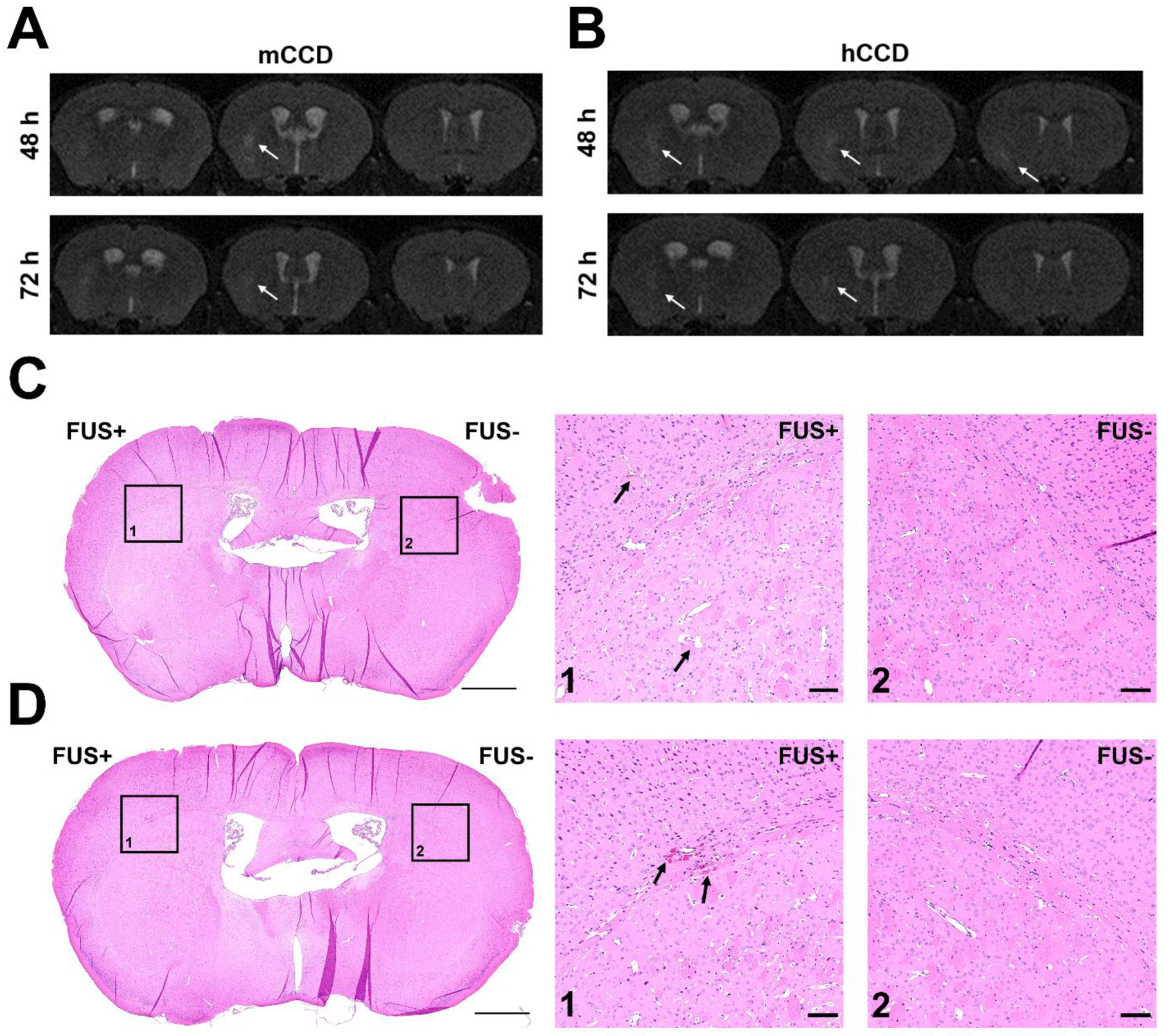
Figure 4.

Consecutive coronal T2-weighted MRI images acquired in mice receiving A) medium CCD or B) high CCD taken at 48 hours and 72 hours after FUS-induced BBB opening. Hyperintense areas indicated by the white arrows represent regions of edema, and were only present in the sonicated hemisphere on the left. No hypointense pixels were visible, indicating a lack of hemorrhaging. Histology using H&E staining was performed, and minimal RBC extravasation was found in only the high CCD group at both C) 6 hours and D) 72 hours after FUS-induced BBB opening. The left striatum was sonicated (FUS+) and the right striatum was used as the control (FUS-). RBC extravasation was only found in the boxed region in the left striatum, with the contralateral side shown as a control. The scale bar for full coronal section view is 1 mm and the scale bar for the zoomed in region is 100 um.
