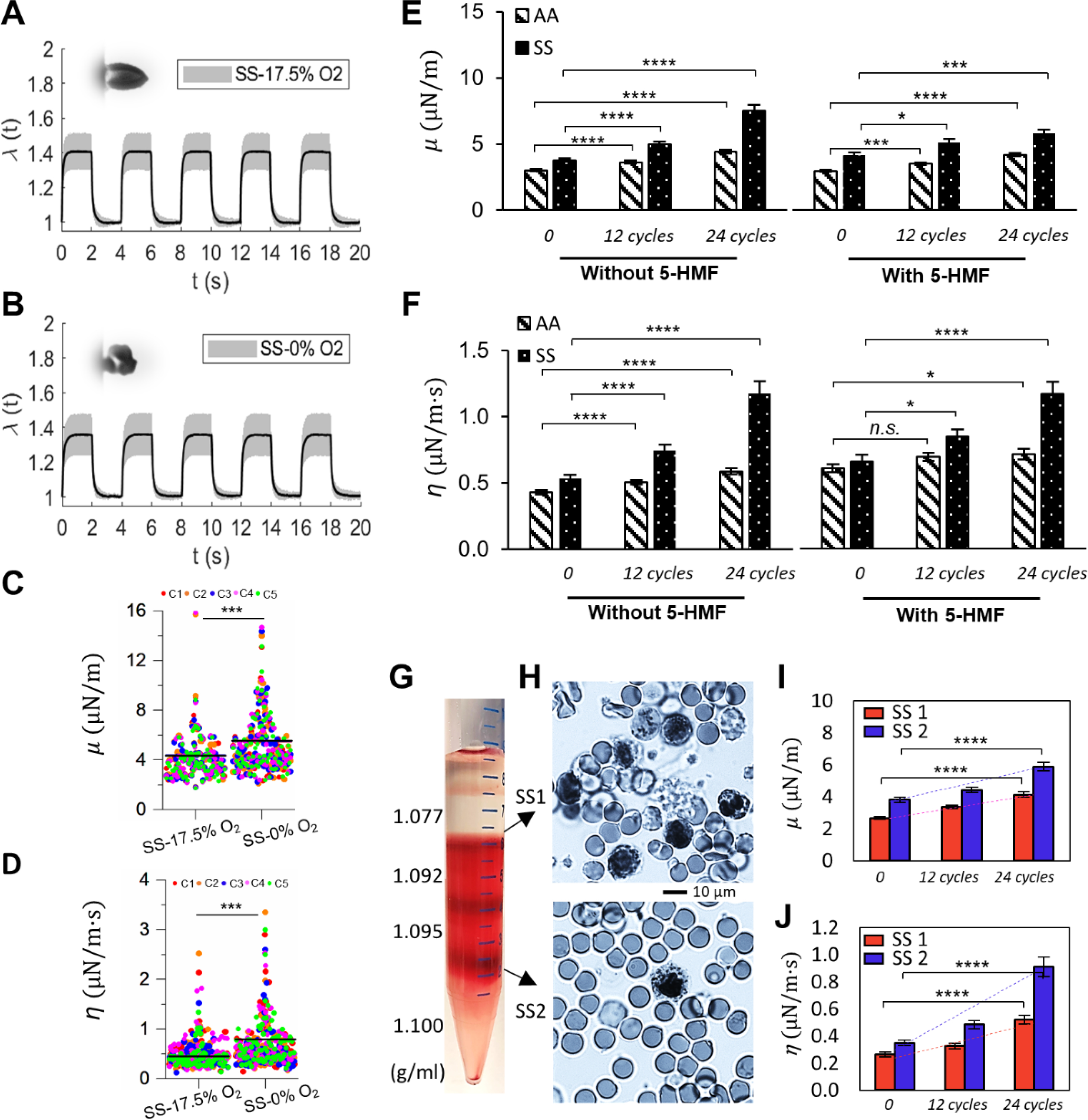
Fig. 5.

Deformability of sickle RBCs (SS) measured under different oxygen conditions. (A and B) Instantaneous values of λ averaged from individually tracked SS cells in the 5 continuous cycles of mechanical testing. Insets show representative images of deformed SS RBCs. (C and D) Comparisons of the corresponding values of μ, and η of normal RBCs under 17.5% O2 and 0% O2. Each symbol represents a single cell measurement. Each color of the data points represents a different measurement cycle in the 5 cycles (C1-C5). (E and F) Comparison of changes in the values of μ and η of AA RBCs and SS RBCs under cyclic DeOxy-Oxy (120s-30s). Data is measured at N = 0 cycle, 12 cycles and 24 cycles. Further comparisons were carried out between samples with treatment and without treatment of 5-HMF. (G) Density separation of RBCs from SCD patients by different cell density fractions of 1.077, 1.092, 1.095 and 1.100 g/mL. (H) Blood smears stained with methylene blue of the reticulocyte-rich (SS1) and erythrocyte-rich (SS2) cell subpopulations. (I and J) Comparison of changes in the values of μ and η of RBCs from SS1 and SS2 sub-populations under cyclic DeOxy-Oxy. * p < 0.05, ** p < 0.01, *** p < 0.001, **** p < 0.001, n.s, not significant. Normal RBCs – AA; Sickle RBCs – SS.
