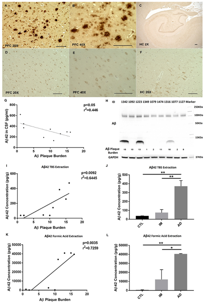
Fig. 1. Aβ and p-tau pathologies of the monkeys.

A-B Aβ plaques in 20X and 40X magnification in prefrontal cortex. C absence of plaque in hippocampus. D-E p-tau in 20X and 40X magnification in prefrontal cortex. F p-tau in hippocampus. Scale bars are 100μm in 20X and 40X images, and 300μm in 2X image. (Abbreviation: PFC Prefrontal Cortex, HC Hippocampus). G relationship between Aβ42 levels in CSF and Aβ plaque burden. Aβ level in CSF is significantly correlated with Aβ plaque burden. H Aβ identified in the PFC of three monkeys with the highest Aβ plaque burden. I relationship between soluble Aβ (TBS extract) levels and Aβ plaque burden. J Levels of soluble Aβ are increased in AD-like group, compared to control or intermediate group. K relationship between insoluble Aβ (formic acid extract) levels and Aβ plaque burden. L Levels of insoluble Aβ are increased in AD-like group, compared to control or intermediate group. Pearson’s correlation analysis was applied. *p<0.05, **p<0.01, one-way ANOVA followed by Tukey’s post hoc test. Note: Two data points superimposed in I and K.
