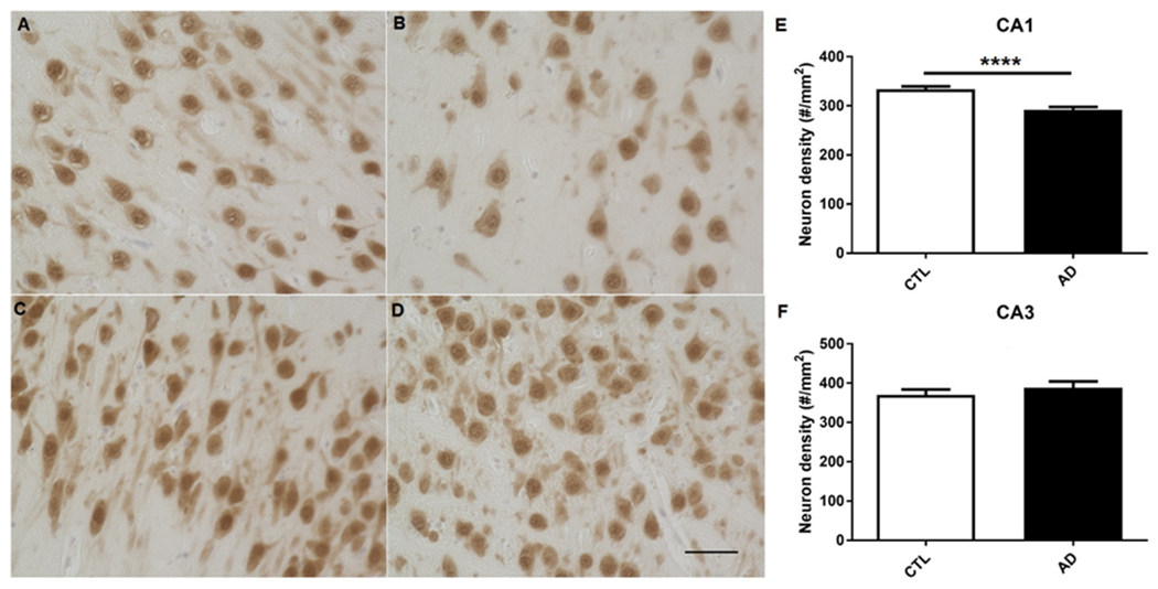
Fig. 4. Neuron density evaluated by NeuN staining in the hippocampi of AD-like and normally aged monkeys.

A representative images of neurons in hippocampal CA1 of normally aged monkeys. B representative image of neurons in hippocampal CA1 of AD-like monkeys. C representative images of neurons in hippocampal CA3 of normally aged monkeys. D representative images of neurons in hippocampal CA3 of AD-like monkeys. E quantification of neuronal density in CA1 of the two groups, indicating neuron density is decreased in CA1 of hippocampi of AD-like monkeys as compared to normally aged monkeys. ****p<0.0001, unpaired independent t test. F quantification of neuronal density in CA3 of the two groups. p>0.05, unpaired independent t test. 40x, Scale bar: 50 μm
