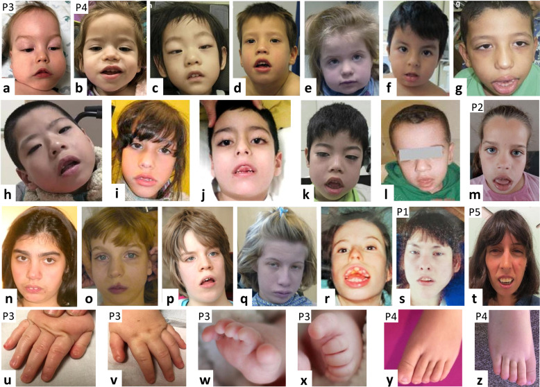
Fig. 1. Facial photographs from 20 individuals with a pathogenic KCNH1 variant.
The facial photos are arranged in order of age from youngest to oldest. The five newly reported patients are indicated by P1–P5. Note the hypotonic facial expression, with open mouth posture and inverted V-shape of the upper lip, and apparent ptosis in some individuals. Facial shape elongates with age (third row), but myopathic facial features remain. a, b Patient 3 (P3; at age 16 months) and patient 4 (P4; at age 1 year 7 months) reported in this study (described in detail in Table 1). c Patient at age 3 years reported in [17] (with permission from Springer Nature). d Patient at age 4 years reported in [48] (with permission from John Wiley and Sons). e Patient at age 4 years 4 months reported in [18] (with permission from Springer Nature). f Patient at age 3 years 7 months reported in [49] (with permission from John Wiley and Sons). g Patient at age 6 years reported in [17] (with permission from Springer Nature). h Patient at age 6 years reported in [17] (with permission from Springer Nature). i Patient at age 6 years 10 months reported in [50] (with permission from Wiley and Sons). j Patient at age 7 years reported in [14]. k Patient at age 8 years reported in [17] (with permission from Springer Nature). l Patient at age 9 years reported in [16]. m Patient 2 (P2; at age 9 years) reported in this study (described in detail in Table 1) and previously reported in [18] (individual 3). n Patient at age 12 years reported in [14]. o Patient at age 13 years reported in [18] (with permission from Springer Nature). p Patient at age 12 years 8 months reported in [14]. q Patient at age 14 years reported in [18] (with permission from Springer Nature). r Patient (age unknown) reported in [14]. s Patient 1 (P1; at age 14 years) reported in this study (described in detail in Table 1). t Patient 5 (P5; at age 34 years) reported in this study (described in detail in Table 1). u, v Fingers of patient 3 (P3; at age 16 months; as described in detail in Table 1), showing proximally placed hypoplastic thumbs with hypoplastic nails. w, x Toes of Patient 3 (P3; at age 14 months; as described in detail in Table 1), showing anonychia of toes 1 and 2 and hypoplastic nails on toes 3–5. y, z Toes of patient 4 (P4; at age 3 years 10 months; as described in detail in Table 1), showing elongated toes with hypoplastic nails.

