
FIGURE 3.
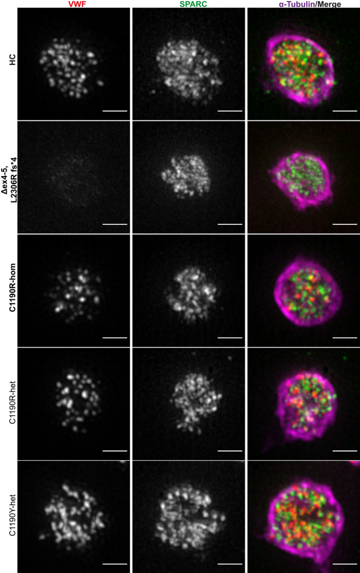
Detailed super‐resolution images of representative platelets from patients with von Willebrand disease (VWD). Platelets from healthy controls (HC), patients with type 3 VWD (Δex4‐5, L2306R fs*4; C1190R‐hom; in bold) and type 2A VWD (C1190R‐het, C1190Y‐het) were stained for VWF (red), SPARC (green), and α‐tubulin (magenta) and were imaged through structured illumination microscopy. Scale bar is 1 µm