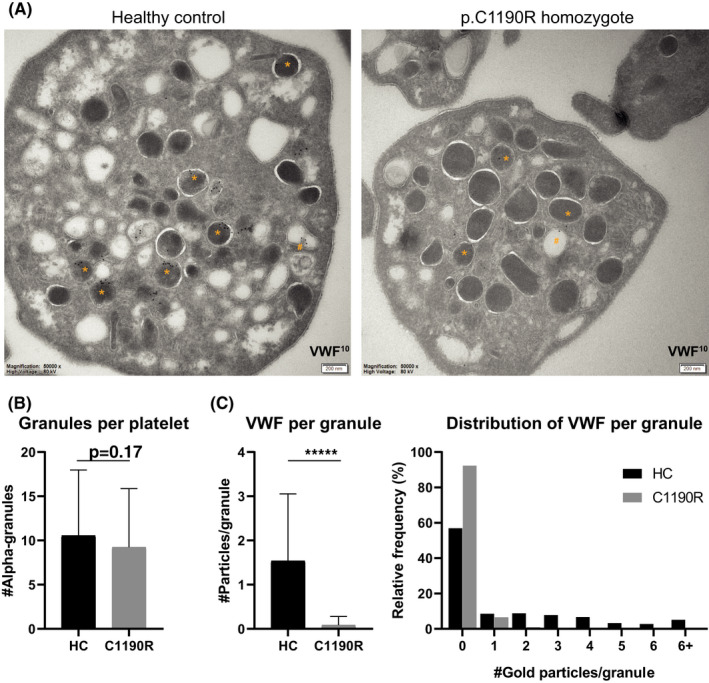
FIGURE 5.

Morphological evaluation of α‐granules and von Willebrand factor (VWF) localization in the p.C1190R homozygous patient. Platelets from a healthy control (HC; n = 73) and homozygous p.C1190R patient (C1190R, n = 75) were assessed through immuno‐electron microscopy for α‐granule numbers and subcellular localization of VWF (A). Immuno‐gold labeling for VWF is shown in α‐granules (*) and open canalicular system (#). Morphologically identifiable α‐granules were quantified (B) and scored for VWF staining inside these granules (C). Data are represented as mean ± standard deviation. ****P < 0.0001
