FIGURE 5.

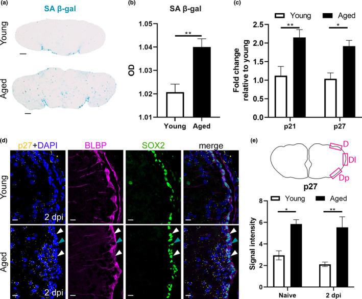
A high senescent cell burden and high expression of cell cycle inhibitors typify the reduced reactive proliferation of progenitor cells in aged killifish. (a) SA β‐gal staining on coronal sections of young adult and aged naive telencephali. (b) Optical density (OD) measurement of SA β‐gal staining of young and aged naive killifish reveals a higher staining intensity in aged killifish, indicating a higher senescent cell burden. Unpaired t test. Values are mean ± SEM; n ≥ 4. (c) Relative expression values of the cell cycle inhibitors p21 and p27 are higher in aged telencephali compared to young telencephali. Two‐way ANOVA is used to compare young and aged fish, followed by Sidak's multiple comparisons test. Values are mean ± SEM; n ≥ 4. (d) Fluorescent in situ labeling of p27 (yellow, HCR), combined with double staining for BLBP (magenta) and SOX2 (green) with DAPI (blue) on coronal sections of young and aged telencephali in naive conditions and at 2 dpi. The expression of cell cycle inhibitor p27 is higher in aged killifish and seems to be localized to RGs (white arrowheads), NGPs (turquoise arrowheads), and newborn neurons (cells lying close to the VZ). (e) p27 signal intensity was measured in a set region of interest in the dorsal (D), dorso‐lateral (Dl), and dorso‐posterior (Dp) portion of the VZ. Aged killifish show a higher signal intensity of p27 compared to young individuals, suggesting increased expression of p27. *p ≤ 0.05, **p ≤ 0.01; unpaired t test is used to compare naive to 2 dpi fish. Two‐way ANOVA is used to compare young and aged fish, followed by Sidak's multiple comparisons test. Values are mean ± SEM; n = 3. SA β‐gal, senescence‐associated β galactosidase; OD, optical density; RG, radial glia; NGP, non‐glial progenitor; VZ, ventricular zone; dpi, days post‐injury; D, dorsal; Dl, dorso‐lateral; Dp, dorso‐posterior
