
Figure 4. Mutual regulation of Plx1 and PP2A‐B56 on Apc1‐loop500 .
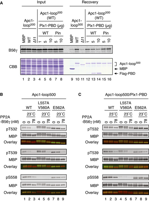
- Plx1 and PP2A‐B56 competes for Apc1‐loop500. MBP‐fused Apc1‐loop500 WT or its derivatives (∆11, deletion of 11 residues including the B56‐binding motif) was incubated with the 35S‐labelled Flag‐B56γ in anaphase extracts supplemented with non‐degradable cyclin B at 23°C for 1 h and further incubated with indicated amounts of WT Plx1‐PBD or Plx1‐PBD Pincer mutant (Pin) for 15 min. The bound proteins were recovered by amylose beads, separated by SDS–PAGE and detected by autoradiography or Coomassie brilliant blue (CBB) staining.
- Dephosphorylation of T532, T539 and S558 on Apc1 by PP2A‐B56. MBP‐fused Apc1‐loop500 WT or its derivatives (L557A/V560A and E562A) was incubated in anaphase extracts supplemented with non‐degradable cyclin B at 23°C for 1 h, isolated by amylose beads and further incubated in the presence (78 nM) or absence of purified PP2A‐B56γ at 23°C for 20 min. The proteins were recovered by amylose beads, separated by SDS–PAGE and detected by immunoblotting with phospho‐specific or MBP antibodies. The signals of a phospho‐specific antibody and MBP antibody on the same membrane were detected in the 800‐ and 700‐nm channels, respectively. The two channels were pseudo‐coloured (green; pT532, pT539 and pS558, red; MBP) and overlaid in the bottom panel (Overlay). The signal of pS558 was very faint in L557A/V560A mutant probably because these two residue are close to S558, interfering with pS558 antibody recognition.
- Dephosphorylation of Apc1‐loop500 by PP2A‐B56 in the presence of Plx1‐PBD. MBP‐fused Apc1‐loop500 WT or its derivatives (L557A/V560A or E562A) was incubated in anaphase extracts supplemented with non‐degradable cyclin B at 23°C for 1 h and further incubated with 10 µg of WT Plx1‐PBD for 15 min. The complexes were isolated by amylose beads and incubated in the presence (78 nM) or absence of purified PP2A‐ B56γ at 23°C for 20 min. The proteins were recovered by amylose beads and analysed as described in (B).