
FIGURE 1.
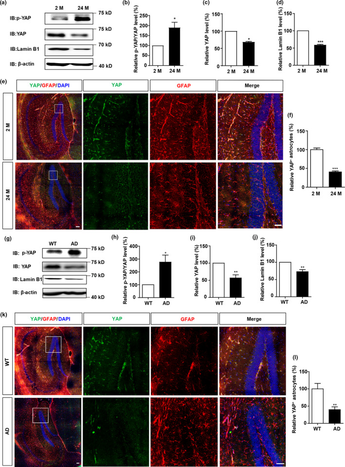
YAP is downregulated and inactivated in hippocampal astrocytes of aged mice and AD model mice. (a) Western blot analysis of p‐YAP, YAP, and Lamin B1 expression in the hippocampus of 2 M and 24 M mice. (b–d) Quantification of relative expression of p‐YAP/YAP (b), YAP (c) and Lamin B1 (d) as shown in (a) (n = 3 per group, normalized to 2 M mice). (e) Double immunostaining analysis of YAP (green) and GFAP (red) in the hippocampus of 2 M and 24 M mice. (f) Quantitative analysis of YAP+ cells over total astrocytes as shown in (e) (n = 3, normalized to 2 M mice). (g) Western blot analysis of p‐YAP, YAP, and Lamin B1 expression in the hippocampus of WT and AD model mice (6 M). (h–j) Quantification of relative expression of p‐YAP/YAP (h), YAP (i) and Lamin B1 (j) as shown in (g) (n = 3, normalized to WT mice). (k) Double immunostaining analysis of YAP (green) and GFAP (red) in the hippocampus of WT and AD model mice (6 M). (l) Quantitative analysis of YAP+ cells over total astrocytes as shown in (k) (n = 3, normalized to WT mice). Scale bar, 100 μm. Data were mean ± s.e.m. *p < 0.05, **p < 0.01, ***p < 0.001