
FIGURE 3.
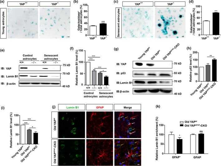
YAP deletion promotes premature senescence of astrocytes in vitro and in vivo. (a) Representative images of SA‐β‐gal staining of young YAP+/+ and YAP−/− astrocytes. (b) Quantification of the percentage of β‐galactosidase+ astrocytes over total astrocytes as shown in (a) (n = 15). (c) Representative images of SA‐β‐gal staining of senescent YAP+/+ and YAP−/− astrocytes induced by D‐gal. (d) Quantification of the percentage of β‐galactosidase+ astrocytes over total astrocytes as shown in (c) (n = 15). (e) Western blot detected YAP and Lamin B1 expression in YAP+/+ and YAP−/− control astrocytes and senescent astrocytes induced by D‐gal. (f) Quantification of relative Lamin B1 expression as shown in (e) (n = 3 per group, normalized to control YAP+/+ astrocytes). (g) Western blot analysis of YAP, p53 and Lamin B1 protein expression in the hippocampus of young YAPf/f mice (2 M), old YAPf/f mice (18 M), and old YAPGFAP‐CKO mice (18 M). (h,i) Quantification of relative expression of p53 and Lamin B1 as shown in (g) (n = 3 per group, normalized to young YAPf/f mice). (j) Double immunostaining analysis of Lamin B1 (green) and GFAP (red) in the hippocampus of old YAPf/f mice (18 M), and old YAPGFAP‐CKO mice (18 M), respectively. (k) Quantitative analysis of the relative average intensity of Lamin B1 as shown in (j) (n = 15, normalized to old YAPf/f mice). ns, not significant. Scale bar, 20 μm. Data were mean ± s.e.m. *p < 0.05, **p < 0.01, ***p < 0.001