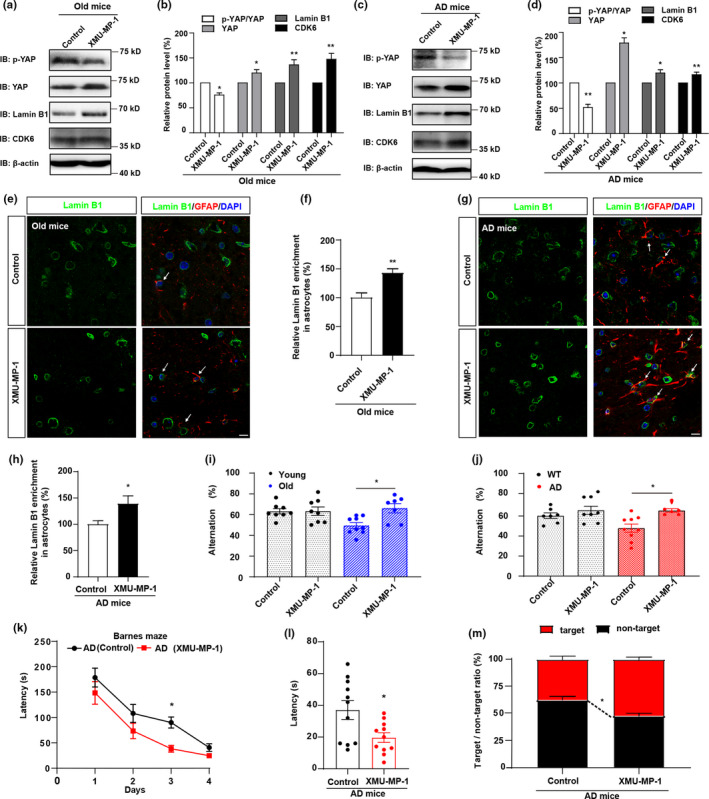
FIGURE 6.

Activation of YAP by suppressing the Hippo pathway improves the cognitive function of aged mice and AD model mice. (a) Western blot analysis of p‐YAP, YAP, CDK6 and Lamin B1 protein expression in the hippocampus of old mice (18 M) with or without XMU‐MP‐1 treatment. (b) Quantification of the relative expression of p‐YAP/YAP, YAP, Lamin B1 and CDK6 as shown in (a) (n = 3 per group, normalized to control treatment). (c) Western blot analysis of p‐YAP, YAP, CDK6 and Lamin B1 protein expression in the hippocampus of AD model mice (6 M) with or without XMU‐MP‐1 treatment. (d) Quantification of the relative expression of p‐YAP/YAP, YAP, Lamin B1 and CDK6 as shown in (c) (n = 3 per group, normalized to control treatment). (e) Double immunostaining analysis of Lamin B1 (green) and GFAP (red) in the hippocampus of old mice (18 M) with or without XMU‐MP‐1 treatment. (f) Quantification of Lamin B1+ and GFAP+ cells over GFAP+ cells as shown in (e) (n = 10, normalized to control treatment). (g) Double immunostaining analysis of Lamin B1 (green) and GFAP (red) in the hippocampus of AD model mice (6 M) with or without XMU‐MP‐1 treatment. (h) Quantification of Lamin B1+ and GFAP+ cells over GFAP+ cells as shown in (g) (n = 10, normalized to control treatment). (i, j) The spontaneous alternation test conducted in WT mice (2 M) and young mice (2 M) (i), old mice (18 M) and AD model mice (6 M) (j) with or without XMU‐MP‐1 treatment by using the Y‐maze (n = 6 mice per group). (k) The time spent to reach the target exit by AD model mice with or without XMU‐MP‐1 treatment in the Barnes maze test from day 1 to day 4 (n = 6 mice). (l) The time spent to reach the target exit by AD model mice with or without XMU‐MP‐1 treatment in the Barnes maze test at the last day (n = 6 mice). (m) The target/non‐target ratio of AD model mice with or without XMU‐MP‐1 treatment (n = 6). Scale bar, 20 μm. Data were mean ± s.e.m. *p < 0.05, **p < 0.01
