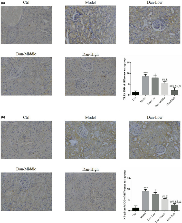
FIGURE 3.

Protein expression of TLR4 and NF‐κB (p65) in renal tissues among the groups by IHC assay (200×). Ctrl: The rats were treated normally; Model: The rats were given DN model treatment; Dan‐Low: DN model rats treated with 10 mg/kg•day Dan; Dan‐Middle: DN model rats treated with 20 mg/kg•day Dan; Dan‐High: DN model rats treated with 100 mg/kg•day Dan. (a) TLR4 protein expression in difference rats groups by IHC assay (200×). ***: p < .001, compared with Ctrl group; #: p < .05, ##: p < .01, ###: p < .001, compared with Model group; $: p < .05, $$: p < .01, compared with Dan‐Low; &: p < .05, compared with Dan‐Middle group. (b) NF‐κB(p65) protein expression in difference rats groups by IHC assay (200×). ***: p < .001, compared with Ctrl group; #: p < .05, ##: p < .01, ###: p < .001, compared with Model group; $: p <.05, $$: p < .01, compared with Dan‐Low; &: p < .05, compared with Dan‐Middle group
