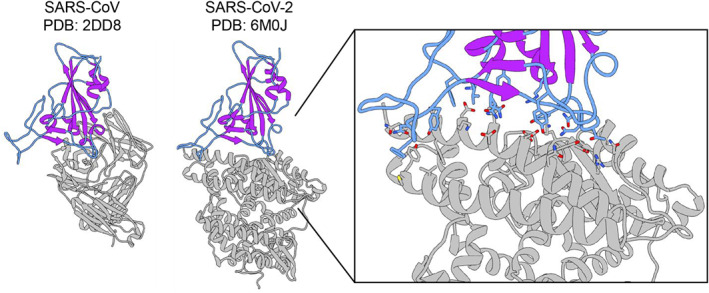
FIGURE 1.

X‐ray crystal structures of CoV1 and CoV2 receptor binding domains (RBD) used in MD simulations. X‐ray crystal structures of the SARS‐CoV RBD in complex with a neutralizing antibody (PDB ID: 2dd8) and the SARS‐CoV2 RBD in complex with the ACE2 receptor (PDB ID: 6m0j). The RBDs from these structures are used as starting structures in this work. The RBD is shown in color and the binding partner is in gray. The loop regions are in blue, and the secondary structure elements are in purple, highlighting the large degree of unstructured regions in the RBD. Enlarged inset of the CoV2 RBD‐ACE2 binding interface shown on the right. Residue sidechains on the RBD (blue) and ACE2 (gray) that participate in the binding interaction are shown in stick configuration
