
FIGURE 1.
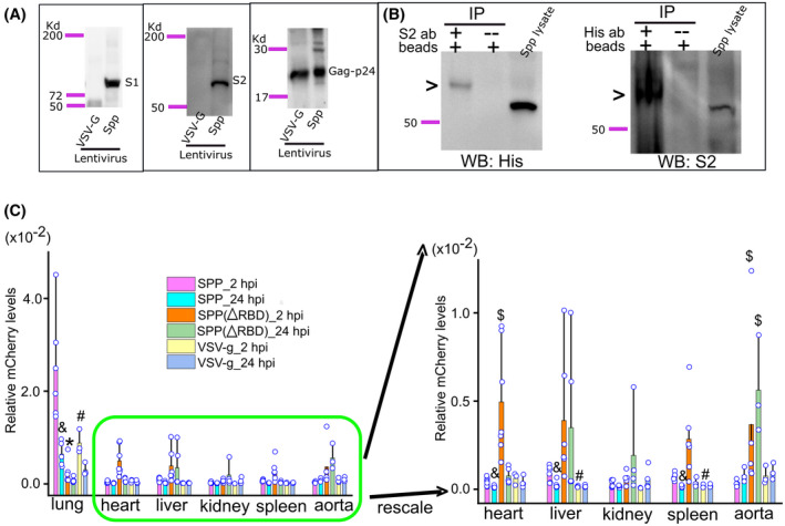
Systemic dissemination of Spp, Spp (∆RBD), and VSV‐G lentiviruses in mice. A, Detection of S1 and S2 subunits in Spp lentivirus by western blot using a specific antibody against the S1 subunit and an anti‐His antibody recognizing the S2 subunit. Both Spp and VSV‐G lentiviruses have a gag‐p24 protein. B, A demonstration of surface S protein on Spp lentivirus. Biotinylation of surface proteins of Spp lentivirus was followed by immunoprecipitation and western blot using an anti‐His or anti‐S2 antibody. A biotinylated S2 protein, indicating by a high molecular weight band by arrows, was detected from immunoprecipitated complexes. C, Spp group shows a predominant distribution in the lungs after intravenous administration. &, P < .05, Spp lentivirus viral burden as compared with VSV‐g lentivirus in the same tissue at 2 hpi; #, P < .05, Spp lentivirus viral burden at 24 hpi as compared with 2 hpi in the same tissue; $, P < .05, Spp (∆RBD) lentivirus viral burden as compared with the Spp group in the heart or aorta at 2 or 24 hpi