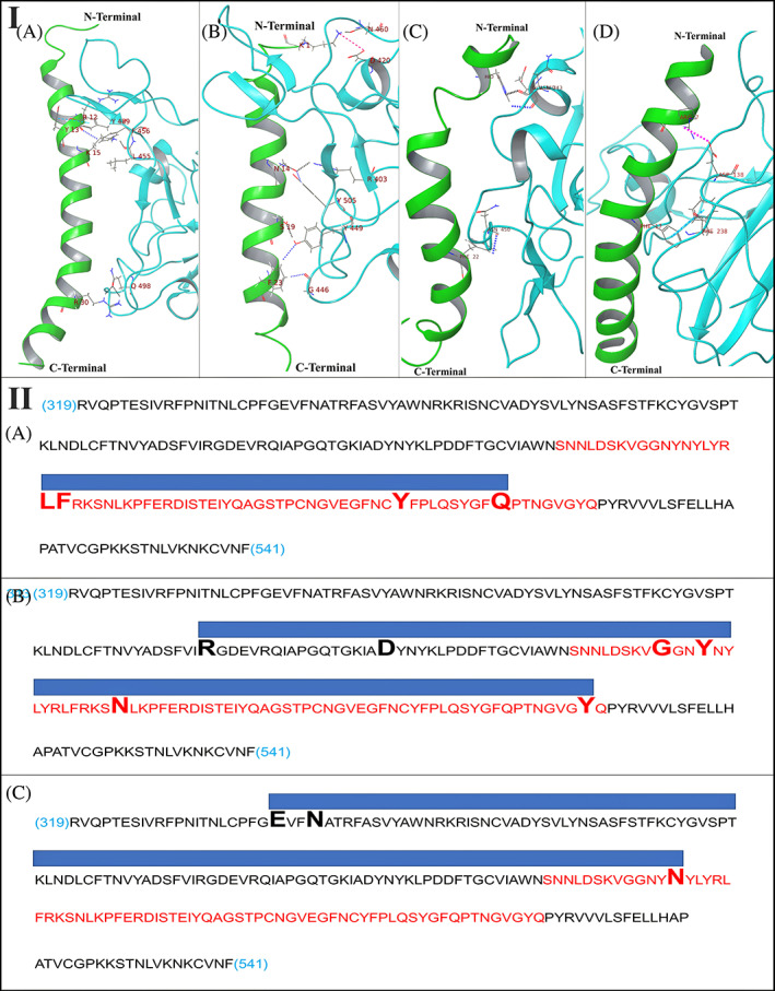
FIGURE 4.

Panel I: Intermolecular interactions and binding mode of (A) PACAP‐38 with SARS‐CoV‐2 RBD, (B) Amylin with SARS‐CoV‐2 RBD, (C) GLP‐2 with SARS‐CoV‐2 RBD, and (D) LL‐37 with SARS‐CoV‐2 region (96–318) which is adjacent to RBD. Hydrogen bonds, aromatic hydrogen bonds, Pi‐Pi stacking and salt bridges are represented by dashed black, blue, sky blue and pink colored lines respectively. Panel II: Diagrammatic representation of molecular docking of control peptides to RBD. Sequence: RBD sequence (319 to 541 of UniProt ID PODTC2 common to 6MOJ and 6LZG); Red sequence: RBM sequence; Large font bold residues: Contacted by the peptide; Blue bar: Contact region covered by the peptide. (A) PACAP‐38 binding to RBD OF 6LZG (Control 1); (B) Amylin binding to RBD OF 6LZG (Control 2); (C) GLP‐2 binding to RBD of 6LZG (Control 3)
