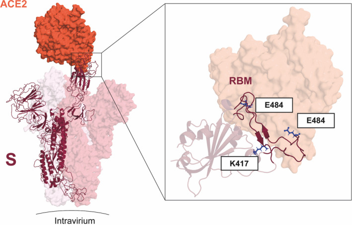
FIGURE 1.

Key mutations of variants B.1.351 and P1 fall on the interface between the RBD and ACE2. (left) S monomer (purple ribbon) bound to ACE2 ectodomain (orange surface). In detail, the positions of residues K417, E484, and N501 (blue sticks) are highlighted. Mutations E484K and N501K occur on the RBM segments (dark purple ribbon), while K417N occurs on helix α4 of RBD. From PDB files 6ACG and 6M0J
