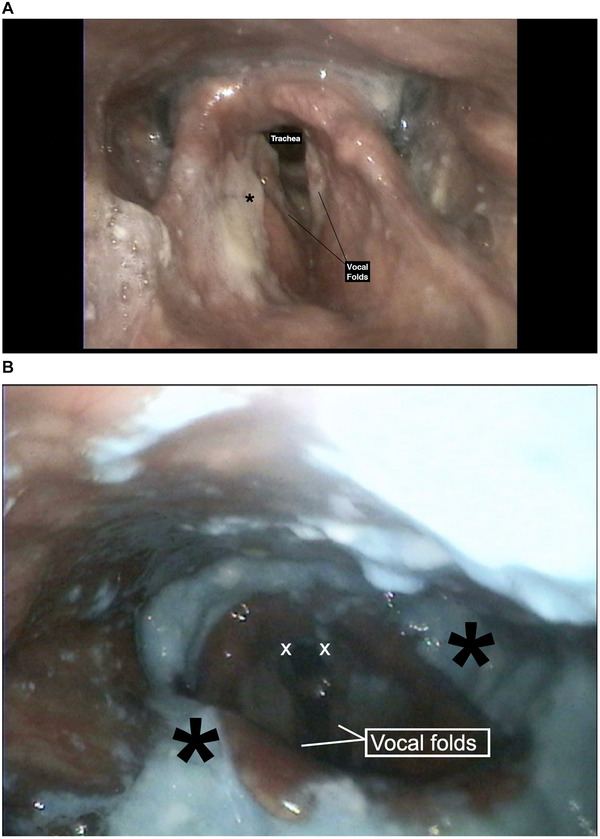
FIGURE 1.

(A) Laryngoscopic evaluation showing laryngeal mucus that does not trigger cough. Black asterisk: Thick mucus over ventricular folds. (B) The presence of residue in valleculae and over the epiglottis with apparent aspiration into the airway. White X: Blue‐dyed food located in the posterior glottis. Black asterisk: Residue collected in the valleculae and pyriform sinuses
