FIG. 8.
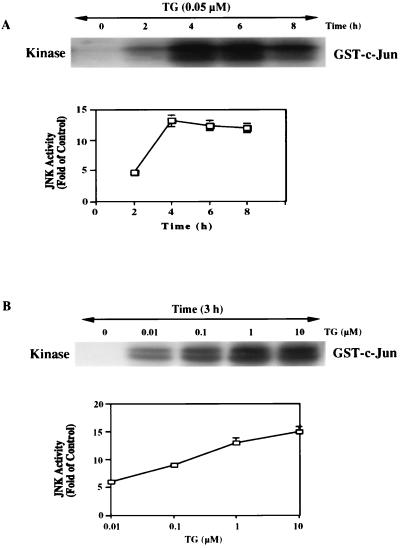
TG induces JNK activation in a time- and dose-dependent manner. Jurkat T lymphocytes were treated with TG for various times. The cell lysates were prepared and immunoprecipitated with 10 μg of polyclonal anti-JNK1 antibody followed by 20 μl of Sepharose A-conjugated protein A. The kinase reaction was performed by the procedures described in Materials and Methods. (A) Time course of the kinase reaction. The top figure represents the autoradiogram of [γ-32P]ATP incorporation into exogenous GST–c-Jun-(1–135). The amount of cell lysate used was 200 μg of protein in each lane. The bottom figure is a plot of JNK activity against time. The values in this figure are means and standard errors of three determinations. (B) Dose response of JNK activation. Anti-JNK1 immunocomplexes were obtained with lysates of cells treated with various doses of TG for 3 h. The JNK assay was performed as described in Materials and Methods. The top figure is a representative autoradiogram of three independent experiments. The bottom figure shows quantified data from three determinations (means and standard errors).

