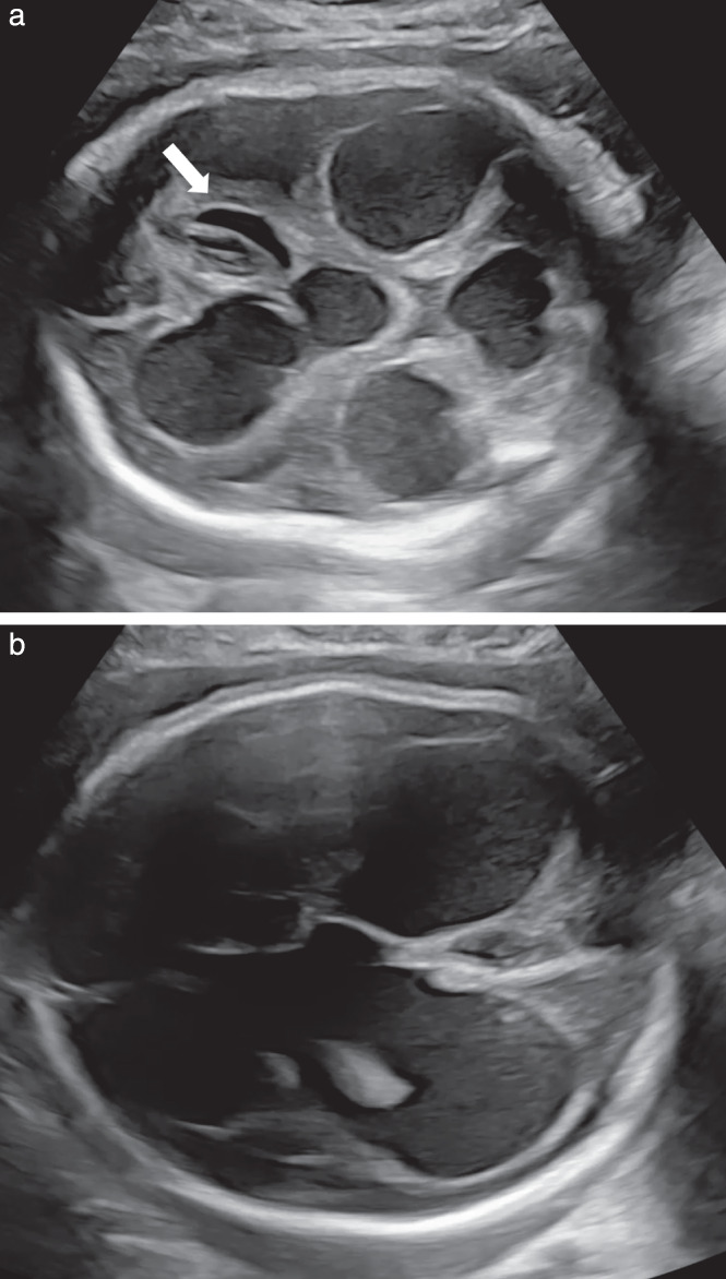
Figure 1.

Grayscale ultrasound images of fetal head obtained at 29 + 4 weeks of gestation in a pregnancy with coronavirus disease 2019, showing progressive hydrocephalus with third and fourth ventricle dilatation, development of porencephalic cysts (arrow) and a disintegrating cerebellum with significant loss of the hemispheres (a) and progressive macrocephaly and hydrocephalus e vacuo (anterior ventricular diameter, 25.9 mm; posterior ventricular diameter 32.8 mm) (b).
