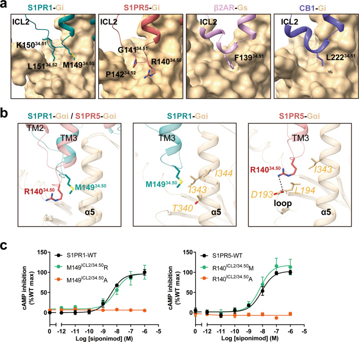
Fig. 6. Role of ICL2 of S1PR1 and S1PR5 in Gi coupling.
a Comparison of contacts between ICL2 regions of the receptors and Gα subunit. ICL2 regions from S1PR1 (teal), S1PR5 (indian red), β2AR (plum) and CB1 (slate blue) are shown as cartoon and Gα subunit from each receptor complex is shown as surface representation. b Structural superposition of ICL2 regions between the S1PR1–Gi complex and S1PR5–Gi complex when aligned on the receptor (left panel). Detailed interactions of M149ICL2/34.50 of S1PR1 with Gαi (middle panel) and R140ICL2/34.50 of S1PR5 with Gαi (right panel) are shown. c Effects of M149ICL2/34.50 mutants of S1PR1 (left) and R140ICL2/34.50 mutants of S1PR5 (right) on siponimod-induced Gi signaling activation potency. Data are presented as the means ± SEM of three independent experiments performed in triplicate.

