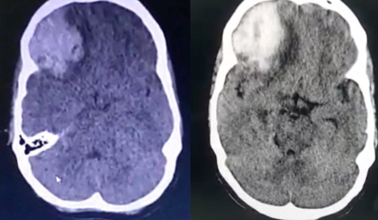
Figure 4.
Non-contrast CT of the brain depicting a large acute intracranial haematoma with surrounding intracerebral oedema in the right frontoparietal cortex with midline shift to left and effacement of basal cisterns suggestive of transtentorial herniation, at admission (left) and 10 days later, at discharge (right), showing reduction of oedema with more clearly defined midline structures.

