Figure 2.
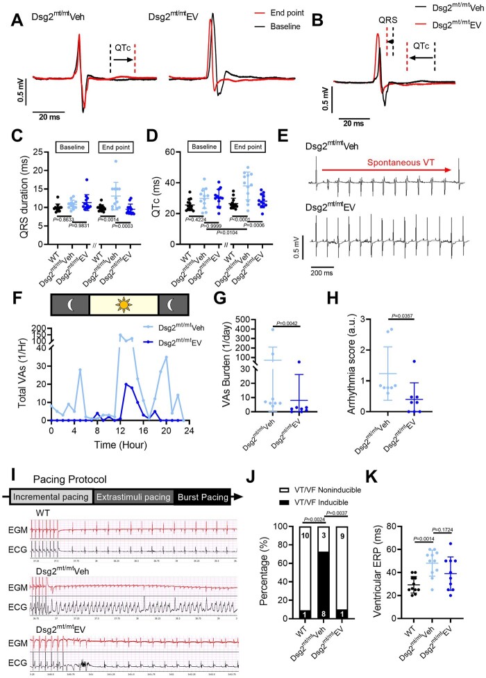
Extracellular vesicles improved ECG abnormalities and ventricular arrhythmias in Dsg2mt/mt mice. (A) Representative electrocardiogram tracings of Dsg2mt/mt mice received vehicle or extracellular vesicles (EVs) highlighting QRS and QT interval changes between baseline and endpoint. (B) Representative electrocardiogram tracings at the endpoint in Dsg2mt/mt mice comparing extracellular vesicle to vehicle group. (C, D) Electrocardiogram parameters of wild type, vehicle-, and extracellular vesicle-treated Dsg2mt/mt mice. Data are mean ± SD (n = wild type: 13; vehicle-injected: 12, extracellular vesicle-treated: 13; one mouse in vehicle group was dead during study period). P-values: one-way ANOVA with the Tukey’s multiple-comparisons test. (E) Representative telemetry electrocardiogram tracings showing ventricular tachycardia (top) in vehicle-injected Dsg2mt/mt mice and a normal electrocardiogram tracing (bottom) in extracellular vesicle-treated Dsg2mt/mt mice. VT, ventricular tachycardia. (F) Twenty-four-hour run chart of total ventricular arrhythmias per hour. VAs, ventricular arrhythmias. (G) Quantification of ventricular arrhythmia burden. (H) Arrhythmia severity score in the vehicle group and the extracellular vesicle group (n = 8 per group). (I) Representative programmed electrical stimulation tracings and pacing protocol (upper panel, intracardiac electrogram; lower panel, surface electrocardiogram) in wild type (top), vehicle-injected (middle), and extracellular vesicle-treated (bottom) Dsg2mt/mt mice. EGM, electrogram. (J) Incidence of pacing-induced ventricular tachycardia (n = wild type: 11, vehicle-injected: 11; extracellular vesicle-treated: 10; one mouse in extracellular vesicle-treated group was dead during study period). (K) Quantification of ventricular effective refractory period (VERP). P-values: one-way ANOVA with the Tukey’s multiple-comparisons test, Mann–Whitney test, and χ2 test.

