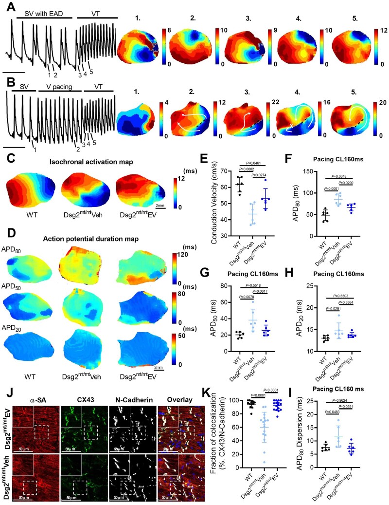
Figure 3.
Mechanisms of ventricular arrhythmias in Dsg2mt/mt mice and electrical remodelling by extracellular vesicle therapy. (A, B) Representative images of optical action potential (left) and isochronal voltages map (right). EAD, early afterdepolarization; SV, sinus ventricular signal; VT, ventricular tachycardia. (C) Representative images of isochronal voltages map and conduction velocity (E). Representative images of action potential duration (APD) map at 160ms pacing cycle length (CL). (F–H) Quantification of action potential duration at 80% (APD80), 50% (APD50), and 20% (APD20) repolarization and action potential duration dispersion (I) in 160 ms pacing conduction velocity (n = 6 per group). (J) Connexin 43 (CX43) immunostaining of the ventricle (upper panel: extracellular vesicle-treated Dsg2mt/mt mice; lower panel: vehicle-injected Dsg2mt/mt mice). (K) Quantification of Connexin 43 and N-cadherin colocalization. Data are mean ± SD (n = 2–3 sections per heart, 6 hearts per group). P-values: one-way ANOVA with the Tukey’s multiple-comparisons test.

