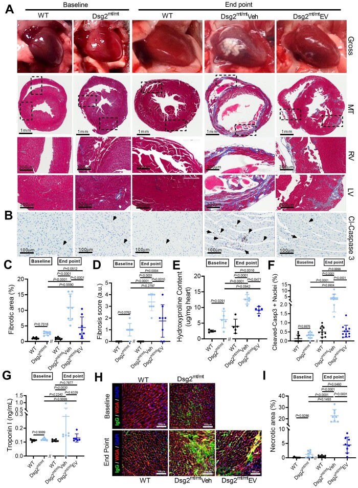
Figure 4.
Extracellular vesicles ameliorate underlying cardiac fibrosis in Dsg2mt/mt mice. (A) Representative gross pathology (top) and Masson’s trichrome-stained micrographs from wild type, Dsg2mt/mt hearts at baseline, and wild type, vehicle-, and extracellular vesicle (EV)-treated Dsg2mt/mt mouse hearts at the study endpoint. Scale bars for ventricle overview: 1 mm; for left and right ventricle: 300 μm. (B) Representative cleaved caspase 3 (cl-caspase 3) immunostaining of apoptotic cells for each group. Scale bars: 100 μm. (C) Pooled data from (A) revealed less fibrosis in extracellular vesicle-treated Dsg2mt/mt mouse hearts (n = 8 per group). (D) Quantification of fibrosis severity (n = 8 per group). (E) Quantification of cardiac hydroxyproline levels (n = 5 per group). (F) Pooled data from (B) revealed less apoptotic cells in extracellular vesicle-treated Dsg2mt/mt mouse hearts (n = 2 sections per heart, 6 hearts per group). (G) Necrosis of cardiomyocytes detected by troponin I release into the serum. (H) Representative images of IgG uptake in wild type, Dsg2mt/mt hearts at baseline, and wild type, vehicle-, and extracellular vesicle-treated Dsg2mt/mt mouse hearts at the study endpoint. Scale bars: 100 μm. (I) Quantification of the extent of cardiac myonecrosis (n = 8 per group). Data are mean ± SD. P-values: Kruskal–Wallis test with the Dunn’s multiple-comparisons test, one-way ANOVA with the Tukey’s multiple-comparisons test.

