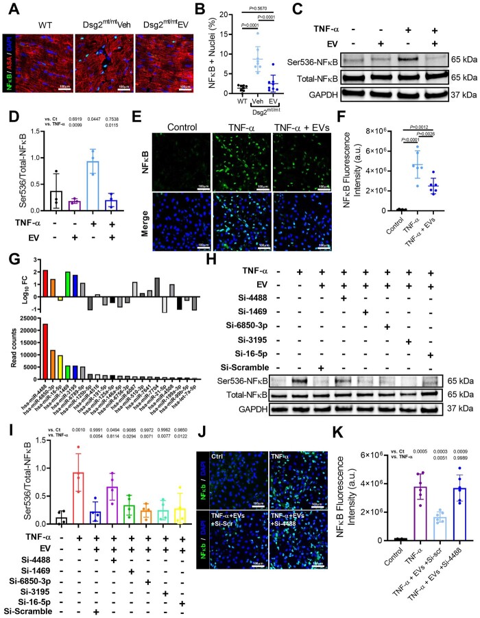
Figure 7.
Reversal of nuclear factor-κB (NF-κB) activation by extracellular vesicles. (A) Immunohistochemical staining for nuclear factor-κB and α-sarcomeric actinin (ASA) in wild type, vehicle-injected, extracellular vesicle-treated Dsg2mt/mt mice. Scale bars: 100 μm. (B) Quantification of per cent of nuclei positive for nuclear factor-κB (n = 8 per group). (C) Representative western blot of neonatal rate ventricular myocyte (NRVM) protein extracts probed with antibodies to phosphorylated and total nuclear factor-κB, and GAPDH in control, TNF-α and extracellular vesicles groups. (D) Quantification of proteins level of phosphorylated and total NF-κB, and GAPDH (n = 3 per condition). (E) Representative immunohistochemical staining from control, NRVMs treated with TNF-α, and NRVMs treated with TNF-α and extracellular vesicles. (F) Quantification of NF-κB fluorescence intensity (n = 6 per condition). (G) Abundance and fold change of top 21 miRNAs identified in the extracellular vesicles. (H) Representative western blot of phosphorylated and total NF-κB, and GAPDH from TNF-α + extracellular vesicle-treated NRVMs transfected with top 5 miR inhibitors. (I) Quantification of proteins level of phosphorylated and total NF-κB, and GAPDH (n = 4 per condition). (J) Representative immunohistochemical staining from control, NRVMs treated with TNF-α, NRVMs treated with TNF-α, extracellular vesicles, and antagomir-scramble, and NRVMs treated with TNF-α, extracellular vesicles, and antagomiR-4488. (K) Quantification of NF-κB fluorescence intensity. (n = 6 per condition). Data are mean ± SD; P-values: one-way ANOVA with the Tukey’s multiple-comparisons test.

