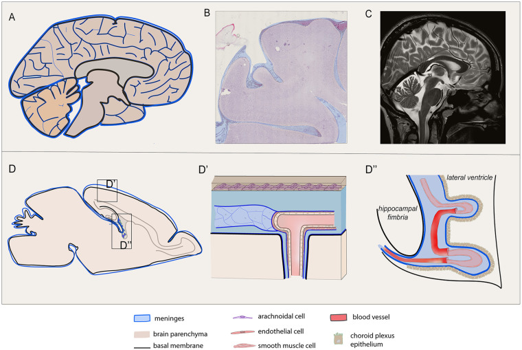
Figure 1.
Meninges are widespread in human and rodent central nervous system (CNS). Meningeal distribution of human (A, B, C) and rodent (D, D′, D′) brain are shown. (A) Sagittal depiction of the human encephalon and (C) the corresponding paramedian T2-weighted magnetic resonance (MR) scan are reported, highlighting the wide distribution of the meningeal layers, excluding the dura mater, as a tissue covering and penetrating inside the cerebral and cerebellar parenchyma, following vessel branches, sulci, and stroma gyration. (B) Coronal section of the human brain stained by hematoxylin and eosin shows meninges penetrating trough the gyri into the sulci. (D) Sagittal graphic view of the rodent brain is reported with enlarged view of the superficial meningeal layer covering the parenchyma at the convexity (D′) and the meningeal substructure penetrating the choroid plexus (D′). (D′) The meningeal arachnoid layer defines the subarachnoid space that is hosting blood vessels, as they deeply penetrate into the sulci and parenchyma in the perivascular spaces projecting through the main brain substructures. The pia mater adheres to the parenchyma and its basal membrane and divides the arteriolar endothelium from the parenchyma. (D′) The pia mater also wraps the choroid plexus (tela choroidea, D′).

