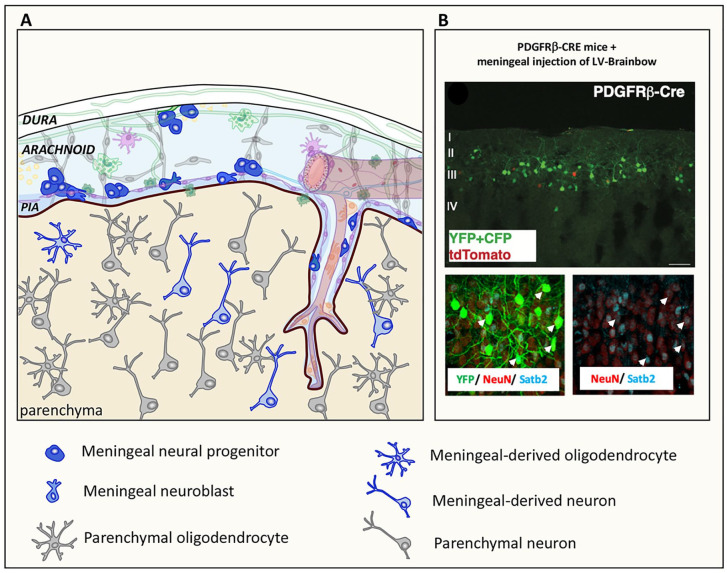
Figure 4.
Meningeal neural progenitors generate parenchymal neurons and oligodendrocytes in physiological condition. (A) Schematic representation showing that immature neural progenitor cells and neuroblasts in meninges generate meningeal-derived neurons or rare oligodendrocytes in the brain parenchyma. In (B) meningeal-derived neurons (YFP+/CFP+, green and tdTomato, red) in the brain cortex of a postnatal day 30 (P30) PDGFRβ-Cre mouse (upper panel) expressing the neuronal markers NeuN and Satb2 (lower panel, arrowhead) are shown. Meningeal cells were labelled by injecting PDGFRβ-Cre P0 mice with a lentiviral vector expressing the Brainbow 1.0(L) reporter in the meninges allowing to trace the Cre expressing PDGFRβ meningeal cells (YFP+/CFP+ cells, green). tdTomato cells (red) are meningeal derived cells that do not express PDGFRβ. The upper panel shows that the meningeal cells migrated into cortical layers II to IV were mostly PDGFRβ-Cre-derived YFP+/CFP+ cells (green). In the lower panel, YFP/CFP meningeal-derived cells (green), NeuN (red), and Satb2 (blue), showing that the PDGFRβ-Cre-derived YFP+/CFP+ cells were NeuN+/Satb2+ neurons (arrows). Modified from Bifari and others (2017).

