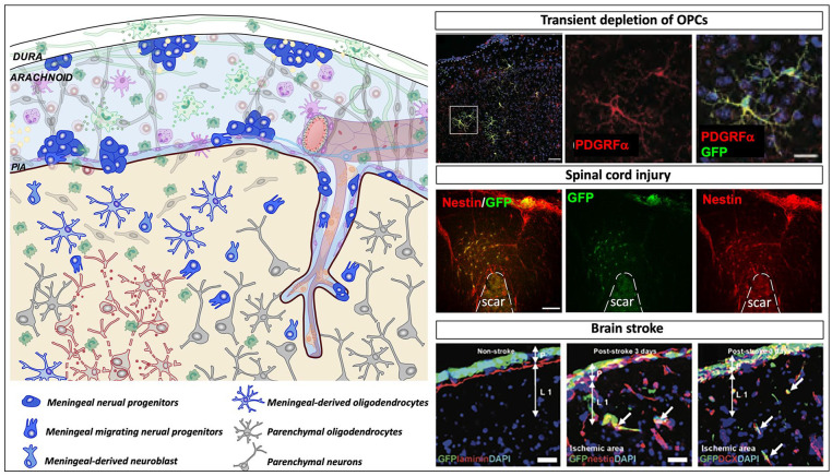
Figure 6.
Meningeal neural progenitors in diseases. Schematic representation showing the meningeal environment in the central nervous system pathological conditions (left panel). Meninges are formed by three tissue membranes: dura mater, arachnoid and pia mater. Following diseases, different signals that include tissue damage, vascular and blood perfusion impairment, cell death, and inflammatory signals activate the meningeal niche. Meningeal progenitor cells, promptly react, proliferate, and migrate from the meninges to the brain parenchyma and differentiate into immature neurons and functional cortical oligodendrocytes. In the right panels, the injury induced meningeal-derived neural cell contribution to three different pathological conditions is shown. In the upper right panel, following transient depletion of oligodendrocyte precursor cells (OPCs), meningeal derived OPCs migrate to the injured parenchyma and differentiate into oligodendrocytes; modified from Dang and others (2019). In the middle right panel, following spinal cord injury, meningeal neural precursors (nestin+, red) migrate to the glial scar site; modified from Decimo and others (2011). In the lower right panel, after brain stroke, meninges increase the expression of nestin- and DCX-positive cells, which migrate to the injured cortex and potentially contribute to cortical regeneration/repair; modified from Nakagomi and others (2012).

