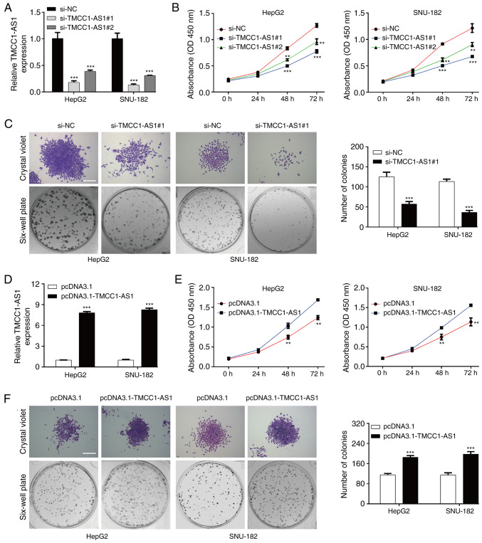
Figure 3.
TMCC1-AS1 promotes proliferation in liver cancer cells. (A) RT-qPCR was used to analyze the expression levels of TMCC1-AS1 in HepG2 and SNU-182 cells transfected with si-TMCC1-AS1#1 or si-TMCC1-AS1#2. (B) Cell viability was determined in the aforementioned transfected HepG2 and SNU-182 cells using a CCK-8 assay. (C) Cell proliferation was assessed using a colony formation assay in HepG2 and SNU-182 cells transfected with si-TMCC1-AS1#1. (D) RT-qPCR was used to analyze the expression levels of TMCC1-AS1 in HepG2 and SNU-182 cells transfected with pcDNA3.1-TMCC1-AS1 or pcDNA3.1. (E) Cell viability was determined in the aforementioned transfected HepG2 and SNU-182 cells using a CCK-8 assay. (F) Cell proliferation was assessed using a colony formation assay in HepG2 and SNU-182 cells transfected with pcDNA3.1-TMCC1-AS1. Data are presented as the mean ± SD of three independent experiments in vitro. **P<0.01, ***P<0.001 vs. si-NC or pcDNA3.1. Scale bar, 50 µm. CCK-8, Cell Counting Kit-8; NC, negative control; OD, optical density; RT-qPCR, reverse transcription-quantitative PCR; si, small interfering RNA; TMCC1-AS1, long non-coding RNA transmembrane and coiled-coil domain family 1 antisense RNA 1.

