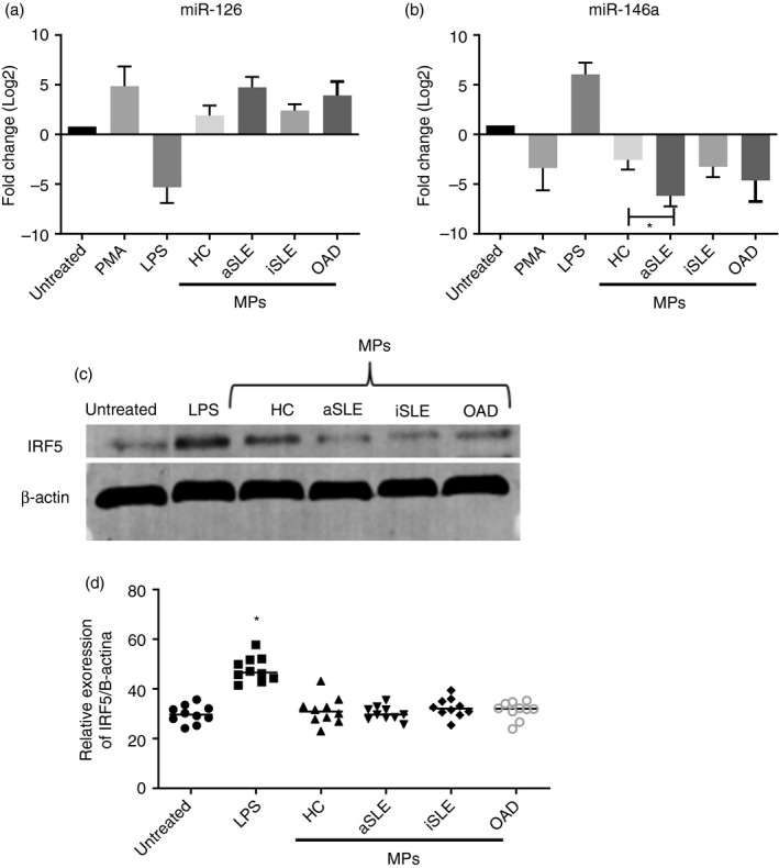
FIGURE 4.

Effect of plasma microparticles on the levels of miR‐126, miR‐146a and IRF5 of U937 cells. U937 cells were untreated or treated for 2 h with plasma MPs (30 µg/µl protein) isolated from healthy controls (HC) and patients. Cell lysates were prepared for miRNA and protein analysis. (a–b) miR‐126 and miR‐146a were amplified by RT‐qPCR, and their quantity was normalised to the relative expression of 18S rRNA. Content of (a) miR‐126 and (b) miR‐146a in cell cultures exposed to MPs isolated from HCs, and patients with active SLE (aSLE), inactive SLE (iSLE) or other autoimmune diseases (OAD). Positive controls: PMA (0·5 µg/µL) and LPS (10 ng/mL). The Kruskal–Wallis test, * p < 0·05. (n = 10 individuals per group). (c) IRF5 expression, evaluated by Western blotting, in cell lysates of U937 cells treated with MPs isolated from HCs and patients with aSLE, iSLE or OAD. Positive control: LPS (10 ng/mL). (d) Densitometry analysis of IRF5 relative to β‐actin. The Kruskal–Wallis test, p < 0·05 (n = 10 individuals per group)
