
FIGURE 5.
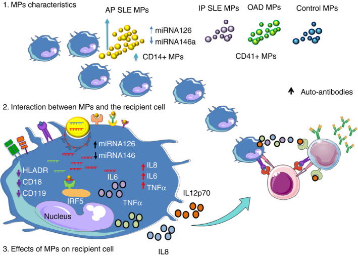
Effect of plasma microparticles of patients with SLE on the expression of miRNAs in phagocytic cells: hypothetical model. 1. Characteristics of microparticles (MPs). Patients with active SLE (aSLE) have a higher concentration of plasma MPs in comparison with patients with inactive SLE (iSLE), other autoimmune diseases (OAD) and healthy controls (HCs). Moreover, aSLE MPs exhibit a higher frequency of monocyte‐derived MPs (CD14+), have higher content of miR‐126 and have lower levels of miR‐146a. The frequency of platelet‐derived MPs (CD41+) is elevated in all study groups. 2. Interaction between MPs and recipient cells. MPs could interact with recipient cells by different mechanisms: linking to cell surface membrane receptors, fusing with cell membranes or undergoing endocytosis/phagocytosis. After, MPs can release their biologically active cargo into the recipient cells. 3. Effect of MPs on recipient cells. Microparticles could mediate intercellular communication, transfer molecular components or induce cellular signalling pathways in the recipient cells. In turn, these events could induce changes in the expression of membrane molecules, cytokine synthesis and/or levels of proteins and miRNAs in the recipient cells. MP‐containing miRNAs induce cellular changes (decreased expression of HLA‐DR, CD18 and CD119, increased synthesis of IL‐8, IL‐6 and TNF‐α, lower levels of miR‐146a, and increasing trend of miR‐126) in recipient U937 cells. However, in comparison with CMPs, aSLE MPs induced significant changes in recipient U937 cells: increased synthesis of IL‐8 and decreased levels of miR‐146a. Therefore, we hypothesized that SLE MPs have particular characteristics that are related to their different effects on recipient cells and could explain their role in the pathogenesis of SLE