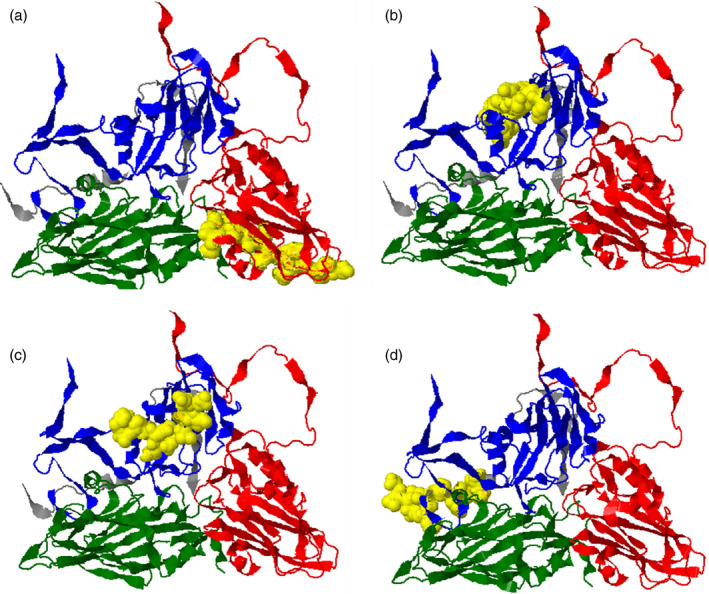
FIGURE 2.

Position of identified epitopes on the protomer of FMDV capsid. Structures (a‐d) are based on the protein data bank: 5nej.1, FMDV‐O1/Manisa model [30] and depict the externally viewed capsid. The structural proteins VP1, VP2, VP3 and VP4 are shown in blue, green, red and grey, respectively. Yellow surface regions indicate the locations of identified epitopes. (a) p220 (VP3: 116‐130), (b) p245 (VP1: 32‐45), (c) p248 (VP1: 46‐60) and (d) p432 (VP4:71‐85)
