
FIGURE 2.
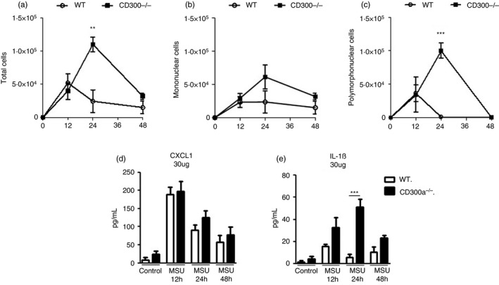
Time course of cell infiltrate and inflammatory mediators in acute gout inflammation. BALB/c and CD300a−/− male mice were injected with MSU crystals into the tibiofemoral joint. Cells from the articular cavity and periarticular tissue were harvested at 12, 24 and 48 hr after injection. From the articular cavity, total cells (a), mononuclear cells (b) and polymorphonuclear cells (c) were analysed. Polymorphonuclear and mononuclear cell populations were determined in cytospin preparation count. Periarticular tissue was processed, and the inflammatory chemokine CXCL1 (d) and the inflammatory cytokine IL‐1β (e) were measured. Bars show the mean ± SEM of 6 mice per group and are from one experiment representative of two independent experiments. Significance was calculated using ANOVA followed by the Newman–Keuls test. **p < 0·005, *** p < 0·001