FIGURE 4.

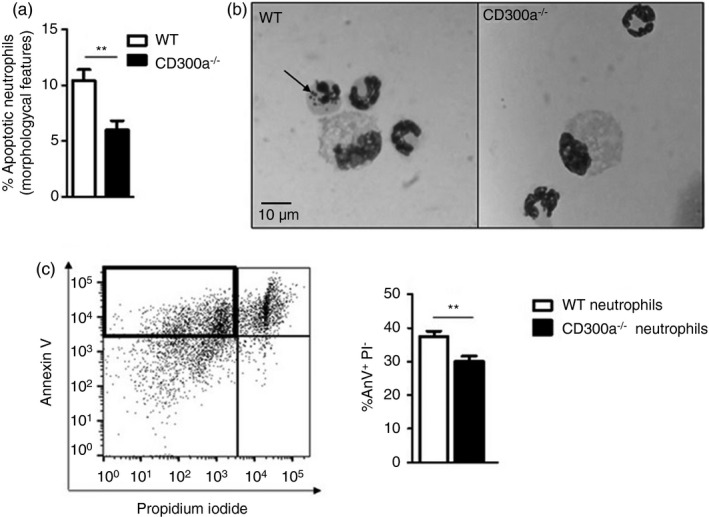
Difference in apoptosis among wild‐type and CD300a−/− neutrophils. BALB/c and CD300a−/− male mice were injected with MSU crystals into the tibiofemoral joint. Cells from the articular cavity were harvested 24 hr after injection, and cells with distinctive apoptotic morphology were evaluated on cytospin and expressed as per cent of neutrophil with apoptotic morphology (a). Representative figures (magnification of 1000×) of viable and apoptotic neutrophil (arrow) (b). BALB/c and CD300a−/− male mice were injected with MSU crystals into the peritoneal cavity. Neutrophils were recovered and isolated 3 hr after peritoneal injection and incubated for 24 hr at 37°. After incubation, neutrophils were stained with annexin V and propidium iodide to assess early apoptosis through flow cytometry (c). The dot plot is an example of general analysis of early apoptosis. Bars show the mean ± SEM of 7 mice per group and are from one experiment representative of two independent experiments. Significance was calculated in relation to the control group (two‐tailed unpaired Student's t‐test). **p < 0·005
