
Fig. 1.
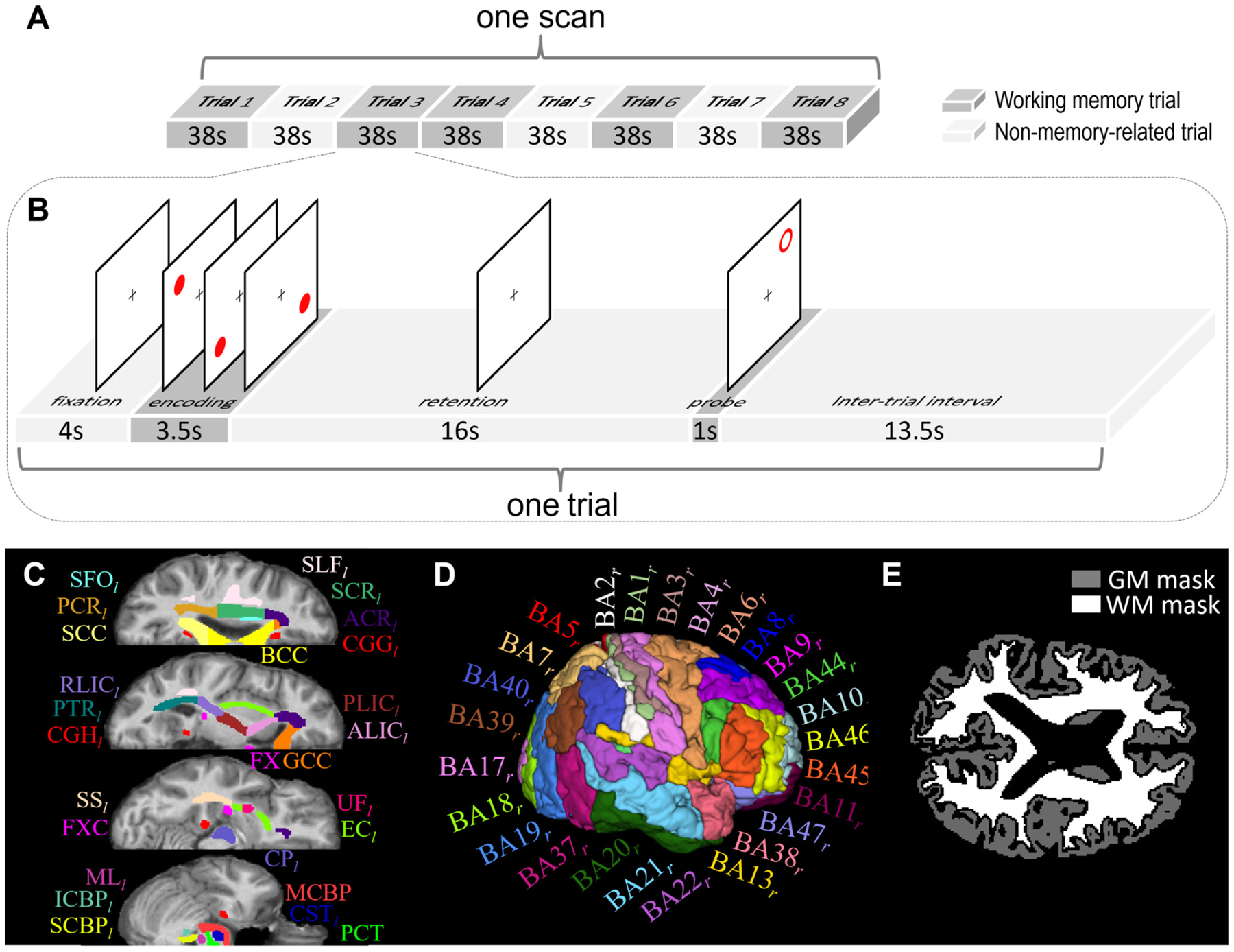
Schematic diagram of spatial working memory task, atlases of white matter (WM)/gray matter (GM) ROIs, and tissue masks. (A) One task scan comprised eight trials, of which five were memory trials (dark gray blocks) and the other three were non-memory trials (light gray blocks). (B) Each memory trial lasted 38s, including 4s of fixation, 3.5s of encoding, 16s of retention, 1s of probe and 13.5s of interval. The non-memory trial had the same sequence of events, except that the subjects were instructed not to remember the target locations. (C) WM parcellation atlas and (D) GM parcellation atlas in MNI space were used to initially define WM and GM ROIs. See Table 2 for the lists of these ROIs. (E) Whole-brain WM and GM tissue masks of one subject that were used to further constrain WM and GM ROIs to avoid partial volume effect.