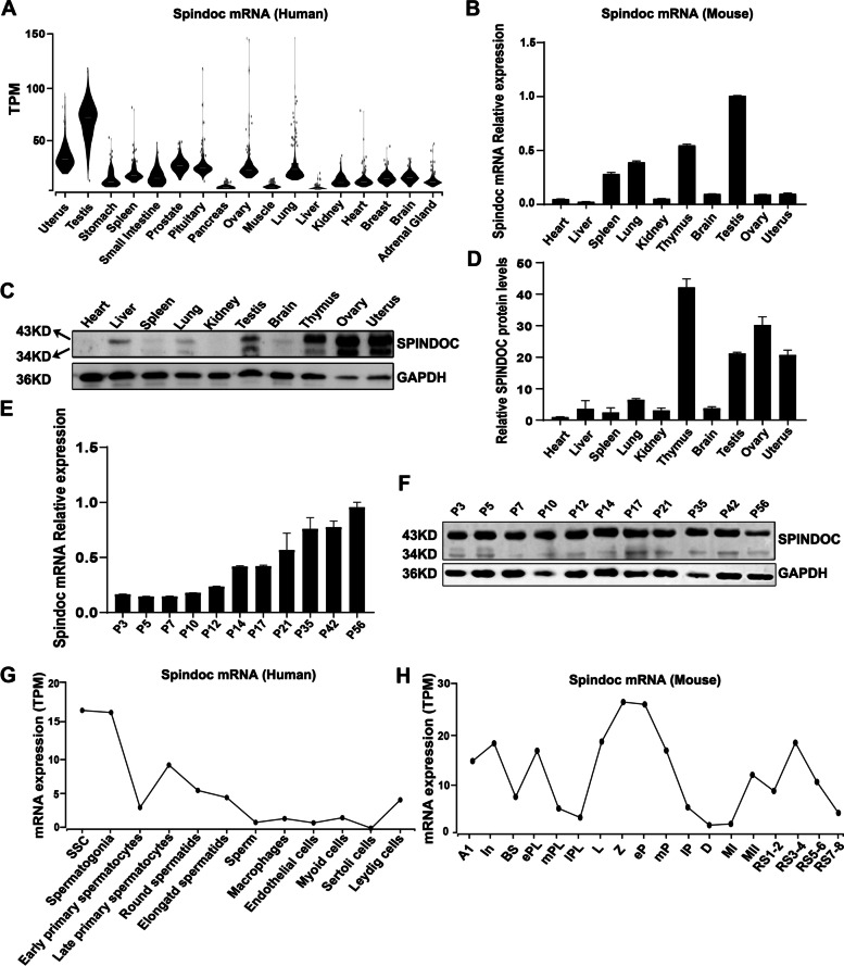
Fig. 1.
Spindoc is predominantly expressed in testis. (A) Violin plots showing the mRNA expression pattern of Spindoc in multiple human organs in the GTEx database (TPM, Transcripts Per Million). (B) RT-qPCR analyses of Spindoc mRNA levels across ten organs from adult wild-type (WT) mice. Data are presented as mean ± SEM, n = 3. (C) Western blot showing the expression levels of Spindoc protein among ten organs of WT adult mice. GAPDH served as a loading control. (D) Densitometric quantification of Spindoc protein levels as in (C). Data are presented as mean ± SEM, n = 3. (E) RT-qPCR analyses of Spindoc mRNA levels in developing testes. Testes at postnatal Day 3 (P3), P5, P7, P10, P12, P14, P17, P21, P35, P42 and P56 were analyzed. Data are presented as mean ± SEM, n = 3. (F) Western blot illustrating the Spindoc protein levels in developing testes. Testes at postnatal Day 3 (P3), P5, P7, P10, P12, P14, P17, P21, P35, P42 and P56 were analyzed. GAPDH served as a loading control. (G) Dynamic expression pattern of Spindoc mRNA from single cell RNA-seq analyses in adult human testes [18]. SSC, Spermatogonial Stem cells. (H) Dynamic expression levels of Spindoc mRNA from single cell RNA-seq analyses in RA-synchronized testicular cells [19]. A1, type A1 spermatogonia; In, intermediate spermatogonia; BS, S phase type B spermatogonia; ePL, early preleptotene; mPL, middle preleptotene; lPL, Late preleptotene; L, leptotene; Z, zygotene; eP, early pachytene; mP, middle pachytene; lP, late pachytene; D, diplotene; MI, metaphase I; MII, metaphase II; RS1–2, steps 1–2 spermatids; RS3–4, steps 3–4 spermatids; RS5–6, steps 5–6 spermatids; RS7–8, steps 7–8 spermatids

