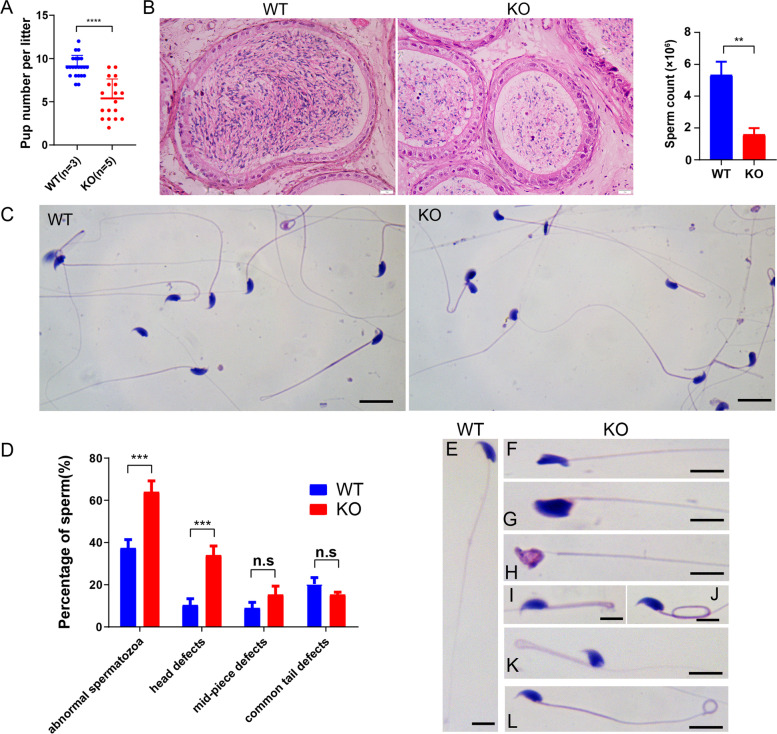
Fig. 3.
Spindoc KO caused impaired sperm production leading to male subfertility. (A) Number of pups per litter from male mice (> 8 weeks) naturally crossed with WT female mice (> 6 weeks) for 4 months. Data are presented as the mean ± SD, n = 5, p < 0.0001 by student t-test. (B) Histological analysis of the cauda epididymis from the WT and KO mice. The average number of sperm released from the cauda in KO mice was less than that in WT mice. Scale bar = 50 μm. The histogram showed the number of sperm retrieved from one cauda epididymis of WT and KO mice. Data were presented as mean ± SEM, n = 3. P < 0.01 by student t-test. (C) Histological analysis showing the morphology of sperm from cauda epididymis in WT and KO mice. Scale bar, 10 μm. (D) The histogram showing the percentage of sperm with abnormal morphology in WT and KO mice at the age of 6 months. Data are presented as mean ± SEM, n = 3. P < 0.001 by student t-test. (E) HE staining of sperm with normal morphology in the WT mice. Sperm with head defects, including irregular shape (F, G, H), coiled mid-piece tail (I, J, K), and tail defect (L), were more frequently observed in the KO mice. Scale bar, 5 μm

