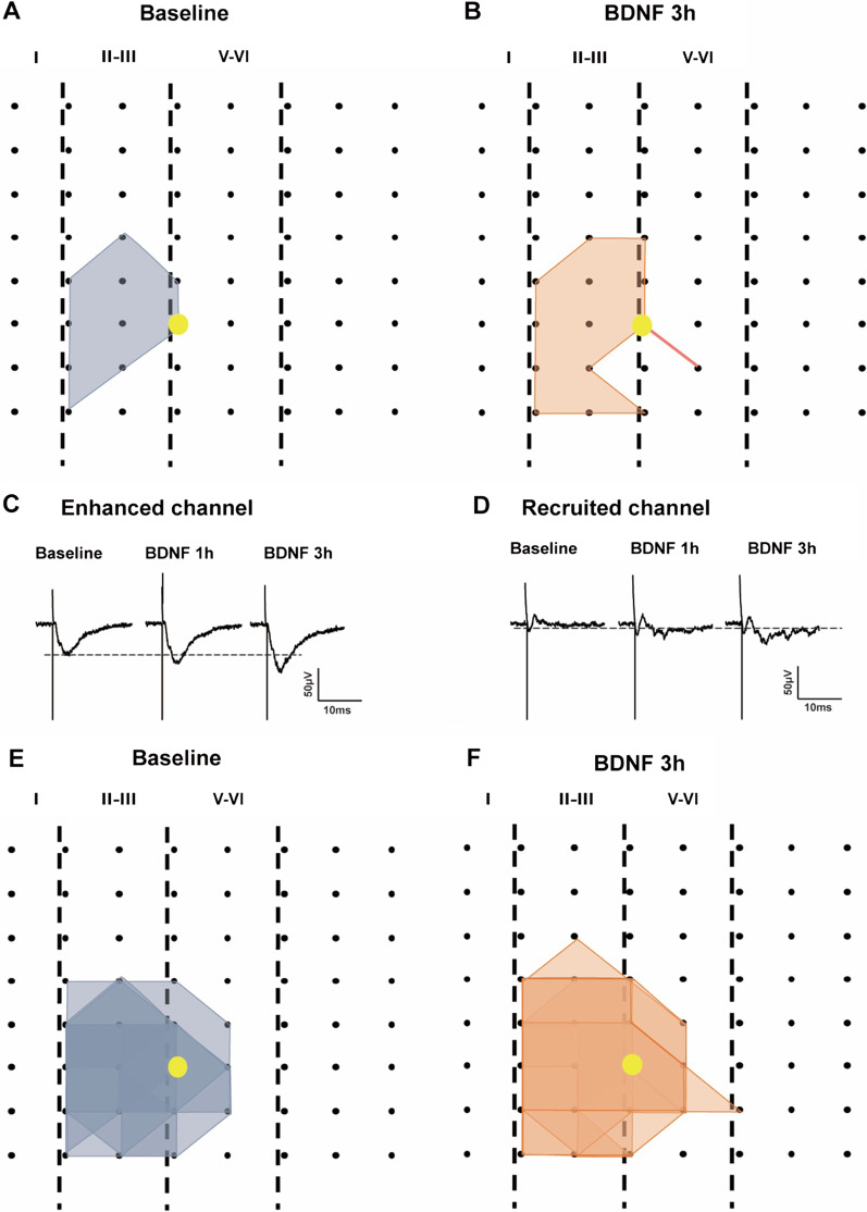
Fig. 3.
BDNF enhanced the network propagation of synaptic responses in the ACC. A, B Sample polygonal diagram showed the distribution of activated channels in the baseline state (gray) and BDNF recruited channels (orange). The yellow circle indicated the stimulation site. C, D The sample traces showed the long-lasting synaptic enhancement and recruited channel at baseline and 1 h, 3 h after BDNF application. E, F Superimposed polygonal diagrams of the activated channels in the baseline state (gray) and the enlarged area after application of BDNF (orange). Black dots represented the 64 channels in the MED64. Vertical lines indicated the layers in the ACC slice

