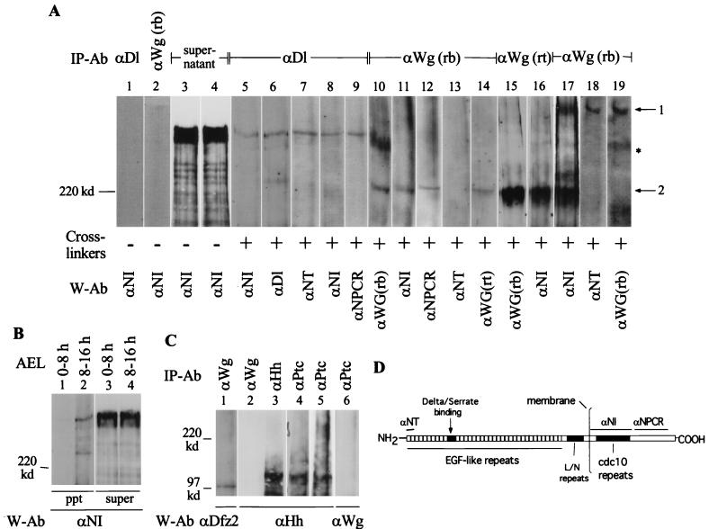
FIG. 4.
Wg and N form complexes during embryogenesis. (A) Two Wg- and N-containing cross-linked complexes, similar to those recovered from cultured cells, are immunoprecipitated from Canton S embryonic extracts. Anti-Dl, is monoclonal antibody MAb 202; anti-NT is described in reference 43; anti-NPCR is described in reference 52; and anti-Wg(rt) was kindly provided by A. Martinez-Arias (See panel D for epitope regions for N antibodies). I used 0- to 3-h embryos for lanes 1 to 16, 6- to 12-h embryos for lane 17, and 10- to 16-h embryos for lanes 18 and 19. Arrow 1 shows Wg complexed with full-length N (lanes 17, 18, and 19); arrow 2 shows Wg complexed with a truncated N (lanes 10 to 17). The asterisk marks the Wg complex not containing N (lanes 10 and 19). A single blot was probed sequentially with the indicated antibodies to form lanes 10 and 11; 12, 13, and 14; 15 and 16; and 19 and 18 (numbers also indicate the sequence of probing). The same embryonic extract was used for lanes 1 to 4 and 7 to 14; lanes 5 to 6, 15, 17, and 18 are derived from different embryonic extracts. (B) Ser-N cross-linked complexes are also recovered from cross-linked embryonic extracts. Complexes were immunoprecipitated with anti-Ser antibody (kindly provided by Elizabeth Knust). Complexes migrating faster than a ∼120-kDa marker protein were not analyzed in panels A and B. (C) The procedure recovering Wg-N, Dl-N, and Ser-N complexes also recovers Wg-Dfz2 (lane 1) and Hh-Ptc complexes from cross-linked embryonic extracts (lanes 3 to 6). For lanes 5 and 6, equal volumes of anti-ptc immunoprecipitate was separated in two different lanes and probed with the indicated antibodies. IP-Ab, immunoprecipitation antibody; cross-linker, BS3; W-Ab, Western blotting antibody; AEL, after egg laying. For panels A and B, 4% polyacrylamide gels were used; for panel C, 6% polyacrylamide gels were used. The tops of all the blots shown in panels A, B, and C coincide with the top of the resolving gel of the discontinuous SDS-PAGE gels. (D) Diagram showing the N epitopes used to produce the N antibodies used in the study.

