FIGURE 4.

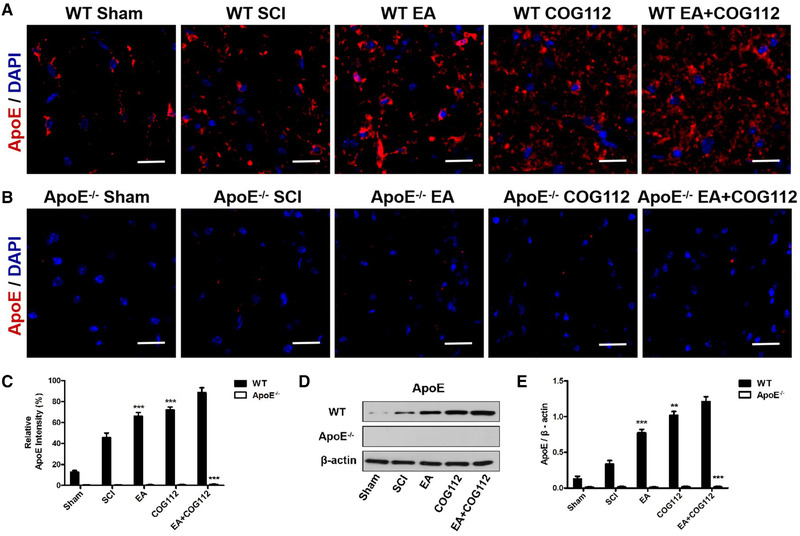
Expression levels of ApoE at 28 d after SCI in spinal cord tissue from WT and ApoE –/– mice (n = 6 samples per group). (a) Immunofluorescence staining of ApoE in WT mice. (b) Immunofluorescence staining of ApoE in ApoE –/– mice. Scale bar = 20 μm. Cell nuclei were stained with DAPI (blue). (c) Quantitative analysis of relative ApoE fluorescence intensity in experiments. (d) Western blot showing the expression of ApoE in the spinal cord from WT and ApoE –/– mice. β‐actin served as the internal control. (e) Quantitative analysis of relative ApoE protein expression in experiments in panel (d). All data are mean ± SD.
Abbreviations: ApoE, apolipoprotein E; DAPI: 4′,6‐diamidino‐2‐phenylindole; EA, electroacupuncture; SCI, spinal cord injury; WT, wild type. ** p < .01; *** p < .001 vs. WT EA+COG112 group
