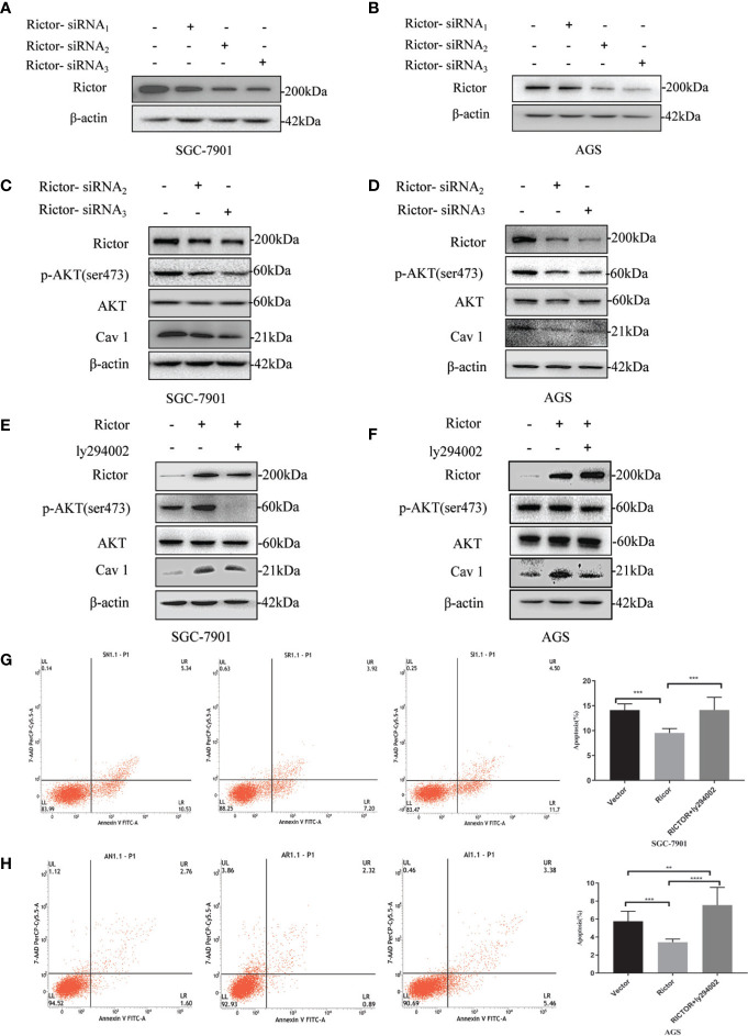
Figure 5.
Association between Rictor and Cav 1 analyzed using western blot and apoptosis detection. (A, B) Two different cell lines were transfected with Rictor-siRNA respectively to verify Rictor-siRNA knock down efficiency. (C) Protein levels of p-Akt and Cav 1 induced by Rictor knockdown in SGC-7901 cells were assessed. (D) Protein levels of p-Akt and Cav 1 induced by Rictor knockdown in AGS cells. (E) After transfection of Rictor plasmid in SGC-7901 cells for 24 h and addition of 20 μM ly294002 for 6 h, the changes of p-Akt and Cav 1 protein levels were detected by western blot. (F) After transfection of Rictor plasmid into AGS cells for 24 h, and addition of 20 μM ly294002 for 6 h, the changes of p-Akt and Cav 1 protein levels were detected by western blot. β-actin served as loading control. (G) Apoptosis of SGC-7901 cells after 24 h transfection with Rictor plasmid and 24 h treatment with 20 μM ly294002 (n=3). (H) Apoptosis of AGS cells after 24 h transfection with Rictor plasmid and 24 h treatment with 20 μM ly294002 (n=3). Values represent the Means ± SD. **P < 0.01, ***P < 0.001 and ****P < 0.0001 were calculated using Student’s t-test.

