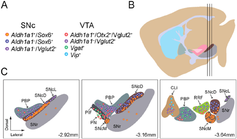
Figure 1.
We outline the regional distribution of seven molecularly defined DAN subtypes in the SNc and VTA: (A) a list of molecularly defined DAN subtypes in the SNc and VTA; (B) a sagittal view of adult mouse brain where the three vertical black lines mark the positions of three cross-sections depicted in (C); and (C) regional distribution of molecularly defined DAN subtypes in the midbrain at Bregma-2.92, −3.16, and −3.64 mm. SNcD: SNc dorsal; SNcM: SNc medial; SNcL: SNc lateral; PBP: parabrachial pigmented nucleus; Cli: caudal linear nucleus of the raphe; PN: paranigral nucleus; PIF: parainterfascicular nucleus; SNc: substantia nigra pars compacta; VTA: ventral tegmental area.

