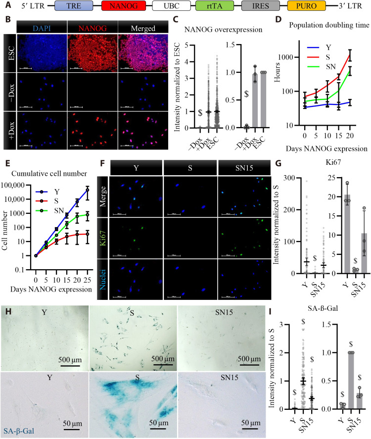
Fig. 1. Overexpression of NANOG in human myoblasts after replicative senescence.
(A) Schematic of the lentiviral vector encoding for NANOG. LTR, Long Terminal Repeat. (B) Immunostaining for NANOG expression and nuclear localization upon addition of doxycycline (Dox) in the medium of NANOG transduced human myoblasts and comparison to endogenous NANOG expression in embryonic stem cells (ESCs). Scale bars, 100 μm. DAPI, 4′,6-diamidino-2-phenylindole. (C) Quantification of fluorescence intensity in (B) reported as means ± 95% confidence interval (CI) for n = 500 cells for each condition in one representative experiment and means ± SD for three donors. (D) Population doubling time for human myoblasts at early passage young (Y), late passage senescent (S), and S myoblasts expressing NANOG (SN), P < 0.05 according to two-way analysis of variance (ANOVA) analysis. (E) Cumulative cell number in Y, S, or SN cells; data shown as means ± SD for n = 3 donors; P < 0.05 according to two-way ANOVA analysis. (F and G) Ki67 immunostaining and quantification for Y, S, and S myoblasts expressing NANOG for 15 days (SN15). Scale bars, 100 μm; data shown as means ± 95% CI for n = 100 cells for each condition in one representative experiment and means ± SD for three donors. (H and I) Senescence-associated β-galactosidase (SA-β-Gal) staining and quantification; data shown as means ± 95% CI for n = 100 cells for each condition in one representative experiment and means ± SD for three donors. $ denotes statistically significant as compared to all other samples.

