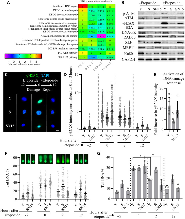
Fig. 4. NANOG improves DNA repair and genomic stability in senescent myoblasts.
(A) GSEA analysis of the DNA repair pathways altered by replicative senescence and NANOG expression for 5, 10, or 15 days. (B) Western blotting analysis of DNA repair proteins as well as phosphorylation of ATM and histone variant H2AX (γH2AX) in response to 2-hour treatment with the genotoxic chemical etoposide. (C) Phosphorylation of histone H2AX (γH2AX) before etoposide treatment (−2 hours) or at 0 and 12 hours after etoposide treatment. Scale bar, 20 μm. (D) Quantification of γH2AX intensity before etoposide treatment (−2 hours) or at 0, 2, and 12 hours after etoposide treatment; data shown as means ± 95% CI for >100 cells in one representative experiment of three independent experiments. (E) Fold increase in the intensity of γH2AX for each condition calculated by dividing the intensity of γH2AX after etoposide treatment to the intensity of γH2AX before the treatment, as a metric for DDR activation. Data shown as means ± SD of five independent experiments, and $ denotes P < 0.05 as compared to all other samples. (F and G) Representative images of single-cell gel electrophoresis assay (Comet assay) quantification of % tail DNA before (−2 hours) or at 0, 2, and 12 hours after etoposide treatment; data shown as means ± 95% CI for >100 cells per condition in one representative experiment (F, scale bar, 40 μm) and means ± SD for three donors (G). * denotes P < 0.05 in the comparison.

