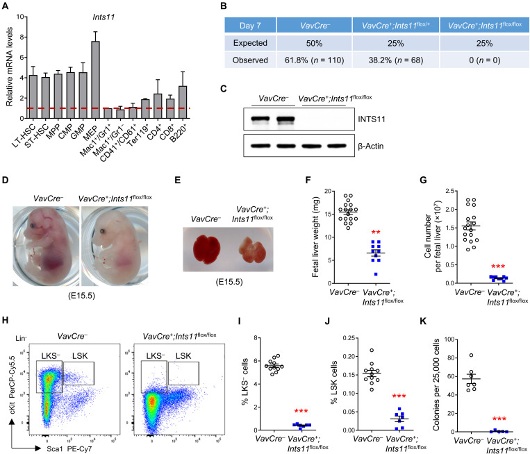
Fig. 1. INTS11 is required for fetal hematopoiesis.
(A) Relative mRNA levels of Ints11 in different hematopoietic cell populations from the BM of WT mice. n = 2 independent experiments; the hematopoietic populations were sorted from five WT mice in each experiment. Long-term HSC (LT-HSC), Lin−Sca1+cKit+CD34−CD135−; short-term HSC (ST-HSC), Lin−Sca1+cKit+CD34+CD135−; multipotent progenitor (MPP), Lin−Sca1+cKit+CD34+CD135+; common myeloid progenitor (CMP), LKS−CD34+CD16/32intermediate; granulocyte-macrophage progenitor (GMP), LKS−CD34+CD16/32high; megakaryocyte-erythroid progenitor (MEP), LKS−CD34−CD16/32low. (B) Genotype distribution in the offspring of the intercross Vav1Cre+;Ints11flox/+ × Vav1Cre−;Ints11flox/flox mice on day 7 after birth. (C) Vav1Cre-mediated INTS11 depletion was verified in fetal liver CD45+ cells from E13.5 Vav1Cre+;Ints11flox/flox embryos. (D and E) Representative embryos (D) and livers (E) from Vav1Cre+;Ints11flox/flox and control littermates at E15.5. Photo credit: Peng Zhang, University of Texas Health Science Center at San Antonio. (F and G) Weight (F) and cellularity (G) of fetal livers from Vav1Cre+;Ints11flox/flox (n = 10) and control littermates (n = 17) at E13.5. (H) Flow cytometric analysis of HSPCs (LSK and LKS−) from control and Vav1Cre+;Ints11flox/flox fetal livers at E13.5. Lineage-negative cells are shown. (I and J) Quantification of the percentages of LKS− (I) and LSK cells (J) from control (n = 11) and Vav1Cre+;Ints11flox/flox (n = 7) fetal livers at E13.5. (K) CFU-C assay using E13.5 fetal liver cells from control (n = 7) and Vav1Cre+;Ints11flox/flox embryos (n = 5) was assessed in semisolid medium in the presence of SCF, IL-3, TPO, GM-CSF, EPO, and IL-6. Data are means ± SEM. Unpaired Student’s t test: **P < 0.01 and ***P < 0.001.

