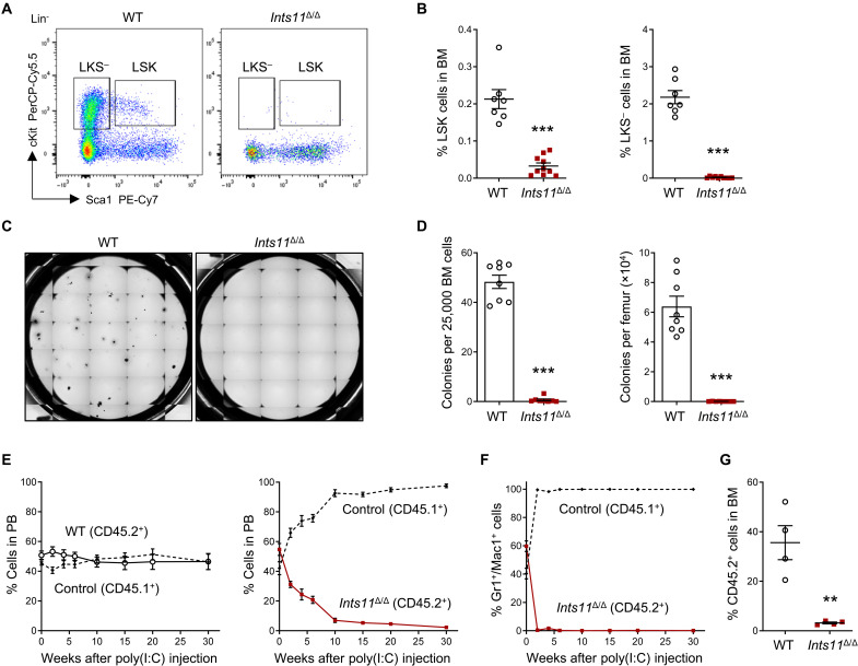
Fig. 3. Loss of Ints11 impairs HSPC development.
(A) Flow cytometric analysis of HSPCs in BM cells from WT and Ints11Δ/Δ mice 12 days after first poly(I:C) injection. (B) Quantification of the percentages of LSK and LKS− cells from WT (n = 7) and Ints11Δ/Δ (n = 10) mice. (C) Representative images of colony formation for Ints11-deficient BM cells and WT controls. The images were taken on the seventh day of the assay. (D) CFU-C assay using BM cells from WT and Ints11Δ/Δ mice (n = 8 per genotype) 12 days after poly(I:C) injection. (E) Percentages of donor-derived (WT or Ints11Δ/Δ CD45.2+) versus CD45.1+ cells in the PB of recipient animals at indicated time points (n = 4 per genotype). (F) Percentages of Ints11Δ/Δ-derived (CD45.2+) versus CD45.1+ cells in the myeloid population of PB of recipient animals at indicated time points (n = 4). (G) Quantification of CD45.2+ cells in BM of recipient animals at 30 weeks after poly(I:C) injection (n = 4 per genotype). Data are means ± SEM. Unpaired Student’s t test: **P < 0.01 and ***P < 0.001.

